/

Everything she covered, taught rather than listed.

The full Lecture Focused guide for neuro, renal and the diabetic emergencies, with every figure and diagram from her slides. Hover any dotted term for its definition; search the glossary from the box up top when an abbreviation stops you.

4 units 19 figures — terms Ch. 61 · 51 · 53 · 17

UNIT 1

Acute Intracranial Problems

Covers: Her 78-slide "Acute Intracranial Problems" deck and the full lecture on it - Lewis's Ch. 61 (pp. 1483-1507) for gaps only

This is the big half of the module. She spent roughly two hours of a two-hour-nine-minute lecture on intracranial pressure and about forty-four minutes on head injury - close to a three-to-one split. This unit is weighted the same way on purpose. Sections 1.1 through 1.14 are increased ICP; 1.15 through 1.18 are head injury and cranial surgery.

1.1 Intracranial Regulation and the Monro-Kellie Doctrine

Everything in this unit follows from one anatomical fact: the skull is a rigid, closed box. Inside it are exactly three things - brain tissue, blood (arterial, venous and capillary), and cerebrospinal fluid (CSF). Because the container cannot expand, the three contents are in permanent competition for a fixed volume.

Intracranial pressure (ICP) is the hydrostatic force measured in the brain's CSF compartment. Balance among the three components is what keeps it normal. Normal ICP is 5-15 mm Hg. A sustained pressure above 20 mm Hg is abnormal and the patient needs treatment. ICP can be measured in the ventricles, the subarachnoid space, the subdural space, the epidural space, or directly in brain tissue with a pressure transducer.

ComponentShare of intracranial volumeWhat it can do when pressure rises
Brain tissue~78% (about 1400 mL)Least able to move. Compensates only by distention of the dura or compression of tissue itself.
Blood (arterial, venous, capillary)~12%Cerebral veins and dural sinuses collapse; regional vessels constrict or dilate; venous outflow changes.
Cerebrospinal fluid~10%Absorption can increase, production can decrease, and CSF can be displaced into the spinal subarachnoid space.

The Monro-Kellie doctrine

The doctrine states that the three components must stay at a relatively constant total volume inside the closed skull. If the volume of one component increases, volume from another must be displaced, and total intracranial volume - and therefore ICP - does not change. That displacement is compensation. It is limited. When the offending volume keeps growing, the other two components run out of room to give, compensation fails, and ICP climbs. Rising ICP compresses tissue, produces ischemia, and ends in cell death.

The three compensatory adaptations

  • Change CSF volume - alter CSF absorption or production, or displace CSF down into the spinal subarachnoid space. This is the fastest and most usable buffer.
  • Change intracranial blood volume - collapse of cerebral veins and dural sinuses, regional vasoconstriction or vasodilation, and changes in venous outflow.
  • Change brain tissue volume - distention of the dura, or compression of brain tissue itself. This is the last and worst option, because compressing brain tissue is the injury you were trying to prevent.

What moves ICP even in a healthy person

Her slide lists six influences as bare words: arterial pressure, venous pressure, intraabdominal and intrathoracic pressure, posture, temperature, and blood gases - especially CO2. In the lecture she gave the mechanism behind three of them, and those mechanisms are what an application question tests.

InfluenceMechanism she gaveBedside consequence
PostureLying flat sends more blood to the head and raises pressure; standing lets gravity pull some of it down.Head-up positioning is an ICP intervention, not a comfort measure.
Intraabdominal / intrathoracic pressureCerebral venous blood drains into the internal jugular veins, then into the inferior vena cava, then to the heart. Pressure on the chest or abdomen presses on the vena cava, so the drainage backs up.Coughing, straining, Valsalva, vomiting, extreme hip flexion and abdominal distention all raise ICP.
TemperatureA higher temperature raises cerebral metabolism; the brain then needs more blood and oxygen; more blood delivered to the head means more pressure.Fever and shivering are treated aggressively (section 1.13).
Blood gases (CO2)Rising PaCO2 relaxes vascular smooth muscle and dilates cerebral vessels.Ventilator settings are an ICP intervention (section 1.2).

1.2 Cerebral Blood Flow, Autoregulation, and CPP

Cerebral blood flow (CBF) is the amount of blood in milliliters passing through 100 g of brain tissue in one minute. Global CBF is about 50 mL/min/100 g. The brain has no fuel reserve, so it needs a constant supply: it consumes 20% of the body's oxygen and 25% of the body's glucose while weighing about two percent of the body.

Autoregulation

Cerebral autoregulation is the brain's automatic adjustment of the diameter of its own blood vessels to hold blood flow constant while systemic blood pressure moves around. Its purpose is to keep CBF consistent and to keep cerebral perfusion pressure within normal limits.

Autoregulation only works across a window of mean arterial pressure. It is effective when MAP is between 70 and 150 mm Hg. Below a MAP of 70, cerebral blood flow falls and symptoms of cerebral ischemia appear - she named syncope and blurred vision, where the slide says only "symptoms of cerebral ischemia." Above a MAP of 150, the vessels are already maximally constricted and have no further response to give; beyond that point, systemic pressure is transmitted straight into the head.

MAP itself is calculated as [SBP + 2(DBP)] / 3. She described it in plain language as "the pressure that keeps the arteries open, allowing sufficient blood flow to the organs and tissues," and gave a practical note: on higher-end monitors the number in parentheses next to the blood pressure is the MAP, so you rarely calculate it

by hand.

Cerebral perfusion pressure

Cerebral perfusion pressure (CPP) is the pressure needed to push blood through the brain. She drew the distinction cleanly: cerebral blood flow is the actual flow going to the cerebrum, and CPP is the pressure required to get that flow through. CPP is what ICP steals.

CPP valueMeaning
60-100 mm HgNormal range.
Below 50 mm HgIschemia and neuron death. (On her slide. She did not read this number aloud - she jumped from "lower CPP causes ischemia" to the under-30 figure - so if the exam tests 50, it is coming from the slide and the chapter.)
Below 30 mm HgIschemia incompatible with life. She did say this one.

CPP = MAP - ICP. The relationship is the whole point of the unit: when ICP rises, CPP falls even if the blood pressure has not moved. That is why maintaining MAP matters so much in a patient with a high ICP, and why lowering ICP is a perfusion intervention rather than a number-chasing exercise. Cerebral vascular resistance - generated by the arterioles inside the cranium - links the two: CPP equals flow times resistance. She explained resistance by analogy to systemic vascular resistance: "what the cerebral arteries are pushing against to get blood flow adequately to the brain tissue."

What CO2 and oxygen do to cerebral vessels

Carbon dioxide, oxygen and hydrogen ion concentration all set cerebral vessel tone. Her framing for the CO2 half was "you may know CO2 is acidic, so anytime there's an increase in PaCO2 you're going to get dilation."

ChangeVessel responseResistanceCBFEffect on ICP
↑ PaCO2Smooth muscle relaxes, vessels dilateRaises ICP - more blood volume in a closed box
↓ PaCO2Vessels constrictLowers ICP, at the cost of perfusion if overdone
Cerebral O2 tension <50 mm HgVessels dilate to recruit flowRaises ICP; the brain is trading pressure for oxygen

She defined cerebral oxygen tension as "the partial pressure of oxygen within the brain tissue - the level of oxygen that's available to the brain cells." If dilation fails to restore it, the tissue switches to anaerobic metabolism, lactic acid accumulates, and the environment turns acidic. Acidosis is itself a potent cerebral vasodilator, so the vessels dilate further in a continued attempt to increase flow. That is the point at which autoregulation is lost and compensatory mechanisms can no longer meet the tissue's metabolic demand. From then on, CBF is dictated directly by systemic blood pressure, hypoxia and catecholamines.

1.3 Increased ICP and Herniation

Increased ICP is a potentially life-threatening state resulting from an increase in any one - or all three - of the components inside the skull. It matters because it decreases CPP and therefore raises the risk of brain ischemia and infarction. The common causes are a mass - hematoma, contusion, abscess, tumor - and cerebral edema.

The cascade - the loop that makes ICP self-worsening

She narrated this entire cascade over a slide that carries no text at all, and it was one of her longest single explanations in the lecture. It is worth learning as a loop rather than a list, because the exam-relevant feature is that each step feeds the step before it.

  • An insult to the brain produces tissue edema.
  • Edema raises ICP.
  • Rising ICP compresses the ventricles and the blood vessels.
  • Compressed vessels mean decreased cerebral blood flow.
  • Decreased flow means decreased oxygen delivery, and brain cells begin to die.
  • Dead tissue draws more edema around the necrotic area - in her words, "once those brain cells die, you're going to get more edema around that necrotic tissue, and that more edema is going to increase the intracranial pressure even more."
  • Higher ICP now compresses the brainstem and the respiratory center.
  • Ventilation fails, so CO2 accumulates.
  • CO2 causes vasodilation, which adds blood volume to a box that is already over-full - ICP rises again, and the loop closes on death.
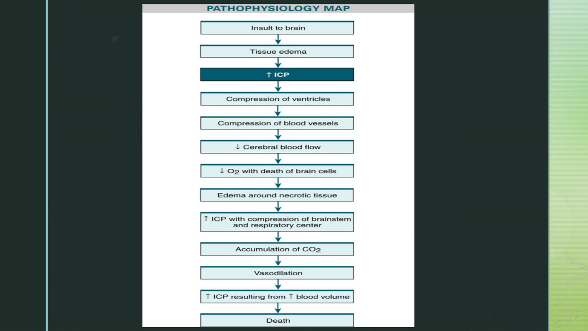
Her pathophysiology map of increased ICP. The slide itself is wordless - this diagram is what she talked over for three minutes. Follow the arrows around: the reason increased ICP is an emergency is that every consequence of it becomes a new cause of it.

Herniation

Herniation is brain tissue being forcibly shifted from an area of greater pressure to an area of less pressure. It converts a potentially reversible problem into an irreversible one, because the shift itself increases ischemia and edema. Herniation forces the cerebellum and brainstem downward through the foramen magnum - which she defined for the class, since the slide only names it: "the hole at the bottom of the skull that allows for the spinal cord to go through."

If the brainstem compression is not relieved, respiratory arrest follows, because the respiratory control center sits in the medulla. With continued herniation, brainstem death is imminent.
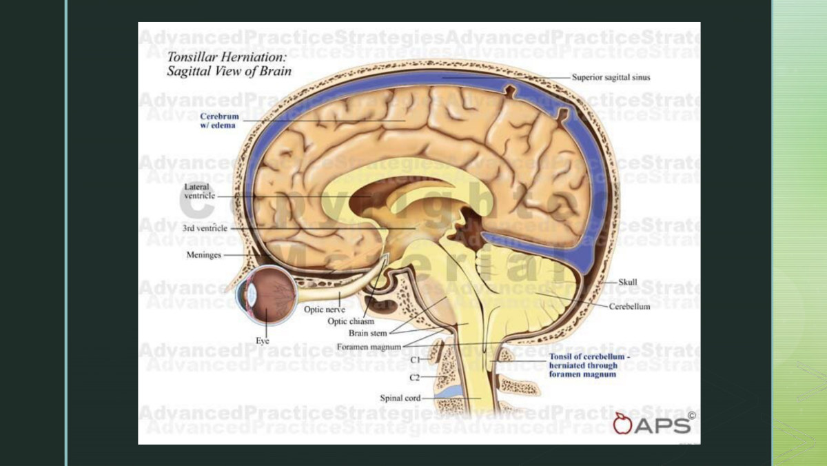
Herniation syndromes - tissue driven across compartments it does not belong in. She narrated the cerebellar tonsil being pushed down through the foramen magnum here. Note that the compartments are separated by folds of dura, not by bone; the dura is what makes the shift damaging.

1.4 The Three Types of Cerebral Edema

Cerebral edema is an increased accumulation of fluid in the extravascular spaces of brain tissue. It raises tissue volume, and therefore raises ICP. The extent and severity of the original insult determine how much edema develops, and a single patient can have more than one type at once.

TypeWhere and why it happensBlood-brain barrierFluid ends up
Vasogenic (most common)Mainly white matter. The blood-brain barrier is disrupted, so large molecules - protein, blood products - enter brain tissue and expose it to toxic products from the blood. That creates an osmotic gradient.DisruptedIntravascular → extravascular space; extracellular fluid volume rises
CytotoxicDisruption of the integrity of the cell membranes themselves, from destructive lesions or trauma to brain tissue - via cerebral hypoxia or anoxia and antidiuretic hormone secretion.IntactExtracellular space → directly into the cells; cells swell and lose function
InterstitialUsually the result of hydrocephalus - fluid building up in the brain, seen as ventricular enlargement. Caused by excess CSF production, obstruction of CSF flow, or inability to reabsorb CSF.Not the mechanismCSF forced out of the ventricles into surrounding tissue

Symptoms of vasogenic edema, and the reporting rule

Vasogenic edema produces headache, decreasing consciousness progressing to coma, and focal neurologic deficits. The slide's warning is that headache can progress quickly to coma and death, so be vigilant. She turned that warning into an actual trigger phrase for calling the provider.

She also named what a focal neurologic deficit actually looks like, where the slide says only the phrase: "speech problems, poor coordination, motor control, changes in personality, or aphasia."

1.5 Manifestations of Increased ICP: Level of Consciousness

Her slide numbers the manifestations 1 through 6, and the order is not arbitrary - it runs roughly from earliest and most sensitive to latest and most ominous. Which manifestations appear depends on the cause of the pressure, its location, and how fast it is rising. A slow-growing tumor and an acute epidural bleed produce the same pressure by very different timelines.

A patient's state of consciousness is defined by clinical responses and by the pattern of brain activity recorded on an electroencephalogram (EEG). Changes can be dramatic - coma - or subtle: a flattening of affect, a change in orientation, a decrease in the level of attention. The subtle end is the dangerous end, because it is the end that gets charted as "patient seems tired."

Coma - the deepest state of unconsciousness

  • No response to painful stimuli.
  • Corneal and pupillary reflexes are absent.
  • The patient cannot swallow or cough - which is the airway problem in section 1.14.
  • Incontinent of urine and stool.
  • EEG shows suppressed or absent neuronal activity.

1.6 Cushing's Triad and the Vital Sign Changes

Changes in vital signs are manifestation number two on her slide, and this is the single hardest-emphasized item in the entire module.

Component of the triadWhat you actually seeWhy it happens
Systolic hypertension with a widening pulse pressureSystolic climbs while diastolic stays low or falls, so the gap between them growsShe gave the mechanism the slide omits: "the reason the systolic blood pressure is increasing is because of the cerebral ischemia. The brain is not getting enough blood flow, so the body is trying to regulate by increasing the systolic blood pressure, trying to get more blood flow to the brain."
Bradycardia with a full and bounding pulseA slow rate you can feel forcefullyBaroreceptor response to the rising pressure, with brainstem cardiovascular centers under compression
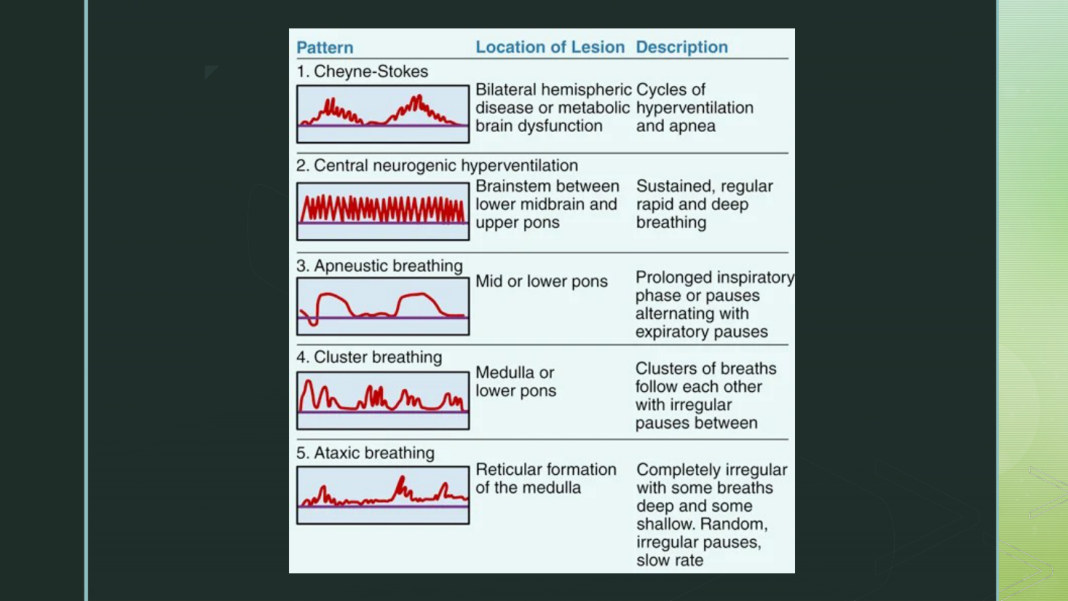
Irregular respirationsRate and pattern both abnormal - see the coma breathing patterns belowDirect compression of the respiratory center in the medulla

The abnormal breathing patterns behind "irregular respirations"

Her deck has two wordless slides of respiratory patterns. She narrated them briefly and pointed students to a video she posted on Canvas of Cheyne-Stokes breathing - a recommendation, not a requirement. The chapter supplies the table itself; it is reproduced here because "irregular respirations" is a category, not a finding.

PatternWhat it sounds likeWhere the lesion is
Cheyne-StokesCycles of hyperventilation alternating with apneaBilateral hemispheric disease or metabolic brain dysfunction
Central neurogenic hyperventilationSustained, regular, rapid and deep breathingBrainstem between the lower midbrain and upper pons
ApneusticProlonged inspiratory pauses alternating with expiratory pausesMid or lower pons
Cluster breathingClusters of breaths with irregular pauses between clustersMedulla or lower pons
AtaxicCompletely irregular - some deep, some shallow, random pauses, slow rateReticular formation of the medulla

These five come from Lewis, not from her slides. What she said about them is the practical instruction: note the respiratory pattern, not only the rate, because specific patterns are associated with severely increased ICP and each one localizes the injury a little lower in the brainstem.

1.7 Ocular Signs of Rising ICP

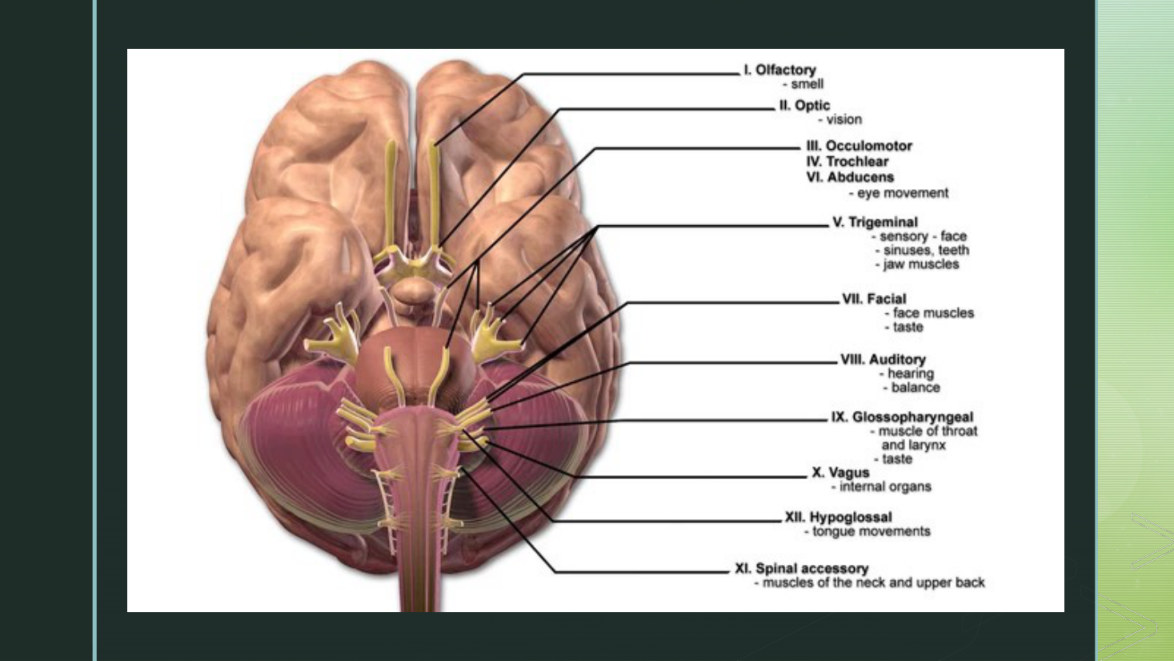
Manifestation number three. Four cranial nerves can be affected - CN II (optic), III (oculomotor), IV (trochlear) and VI (abducens) - but one of them carries almost all of the exam weight.

  • Compression of CN III→ pupil dilation on the same side (ipsilateral) as the mass lesion, a sluggish or absent response to light, inability to move the eye upward and adduct, and ptosis of the eyelid.
  • A fixed, unilateral, dilated pupil is a neurologic emergency indicating brain herniation.
  • If ICP continues to rise, both pupils dilate - the first pupil enlarges until it is fully dilated, then the second follows.
  • CN II, IV and VI problems produce blurred vision, diplopia, and changes in extraocular movements. These are three separate findings; blurred vision and diplopia are not the same thing.
  • Central herniation→ sluggish but equal pupil response.
  • Uncal herniation→ a dilated unilateral pupil.
  • Papilledema - an edematous optic disc - is a nonspecific sign of persistent increased ICP.
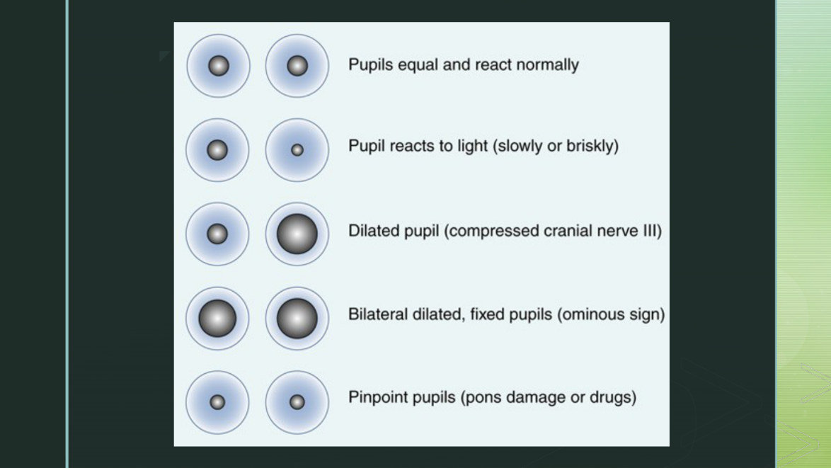
Her pupil findings slide. Read it as a progression rather than a menu: equal and briskly reactive → sluggish on one side (early pressure on CN III) → one pupil dilated and fixed (herniation) → both dilated and fixed.
Her pupil findings slide. Read it as a progression rather than a menu: equal and briskly reactive sluggish on one side (early pressure on CN III) one pupil dilated and fixed (herniation) both dilated and fixed.
The companion pupil illustration. Compare sizes against the millimetre scale rather than eyeballing "big" and "small," and document each pupil separately - the whole diagnostic value here is in the asymmetry.

How pupils are assessed and charted

  • Compare pupils for size, shape, movement and reactivity.
  • A normal reaction is brisk constriction when the light shines into the eye.
  • The consensual response is slight constriction of the opposite pupil at the same time. This is on her slide; she did not say it aloud, so learn it from the deck.
  • A sluggish reaction indicates early pressure on CN III - this is the one to catch.
  • A fixed pupil with no response to light usually indicates increased ICP. The chapter adds other causes that must be ruled out: direct injury to CN III, previous eye surgery, atropine, and mydriatic eye drops.
  • A hand-held pupillometer may be used to remove subjectivity - a chapter addition, not on her slides.

1.8 Motor Findings, Posturing, Headache, and Vomiting

Manifestations four, five and six. Motor change is the one with the localizing value; headache and vomiting are the ones students underweight because they sound benign.

Decrease in motor function

Contralateral hemiparesis or hemiplegia develops - on the side opposite the mass lesion. She isolated the vocabulary deliberately, because the slide uses both words as if they were interchangeable: hemiparesis is weakness or inability to move one side of the body; hemiplegia is partial or complete paralysis. Contralateral means opposite; ipsilateral means same side.

With painful stimuli the patient may localize - move toward the stimulus, which is the better response - or withdraw, moving away. Below withdrawal come the two posturing patterns, which occur because voluntary motor tracts from the cerebral cortex have been interrupted.

Decorticate (flexor)Decerebrate (extensor)
ArmsInternal rotation and adduction, with flexion of elbows, wrists and fingersStiffly extended, adducted and hyperpronated
LegsExtendedHyperextended with plantar flexion of the feet
Where the damage isInterruption of voluntary motor tracts in the cerebral cortexDisruption of motor fibres in the midbrain and brainstem - the chapter supplies this level; her slide says only that it indicates more serious damage
MeaningSeriousMore serious. Deterioration from decorticate to decerebrate is the direction that matters
GCS motor score3 (abnormal flexion)2 (abnormal extension)
Decorticate posturing. Arms drawn in toward the chest, elbows and wrists flexed, legs extended. Her slide prints the memory hook "going to your core"; she expanded it as the patient "protecting their core - everything is going inwards towards the core."
Decerebrate posturing. Arms rigidly extended and rotated palms-outward, legs extended, feet plantar flexed. Everything points away from the body - the opposite picture, and the worse one.

Headache

Headache in increased ICP comes from compression of intracranial structures. The chapter is precise about why: the brain itself has no pain receptors, so the pain is coming from arteries, veins and cranial nerves being squeezed. A nocturnal or early-morning headache is the concerning pattern - it suggests a tumor or another space-occupying lesion, because lying flat overnight raises ICP further. Straining, agitation or movement makes the pain worse, for the same venous-drainage reason from section 1.1.

Vomiting

Vomiting is a nonspecific sign of increased ICP related to the pressure change inside the cranium, and it may be projectile. The chapter adds a discriminator her slide omits: the vomiting of increased ICP is unexpected - not preceded by nausea. That is what separates it from gastrointestinal vomiting in a stem.

1.9 Complications: Inadequate Perfusion and the Herniation Syndromes

The two major complications of increased ICP are inadequate cerebral perfusion and cerebral herniation. Perfusion failure is section 1.2 arriving: as ICP climbs, CPP falls, and below 50 mm Hg neurons start dying. Herniation is the anatomical event.

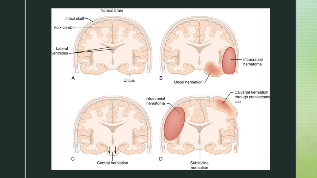
Two structures decide where the tissue can go. The falx cerebri is a thin wall of dura folding down between the hemispheres, separating left from right. The tentorium cerebelli is a rigid fold of dura separating the cerebral hemispheres from the cerebellum - the chapter notes it is named for a tent, because it forms a tent-like cover over the cerebellum. Herniation syndromes are named for which of these edges the brain is pushed across.

HerniationWhat shifts, and whereClue at the bedside
Tentorial (central)A cerebral mass forces the brain downward, toward and through the foramen magnumSluggish but EQUAL pupil response; deteriorating LOC; eventually Cushing's triad and respiratory arrest
UncalLateral and downward herniation of the uncus of the temporal lobe over the tentorial edgeDilated UNILATERAL pupil - CN III compressed on the side of the lesion
Cingulate (subfalcine)Lateral displacement of brain tissue beneath the falx cerebriMay be relatively silent early; the shift is visible on imaging as midline shift
CalvarialHerniation through a craniectomy siteChapter addition - not on her slides. It is the flip side of the craniectomy in 1.18: opening the box relieves pressure but creates a new exit
Her illustration of the herniation syndromes. She framed the slide this way: "these herniation descriptions are just showing where the herniation is occurring - so central, uncal and subfalcine." Match each arrow to the pupil finding in the table above; the pupils are how you tell them apart without a scan.

1.10 Diagnostic Studies and Methods of ICP Monitoring

Diagnostic studies here are used to identify the cause of the increased pressure, not to confirm that pressure is high - the bedside assessment does that. Her list: CT and MRI, EEG, cerebral angiography, ICP measurement, PET, and brain tissue oxygenation measurement with a LICOX catheter.

Who gets an ICP monitor

  • To guide care for any patient at risk for or with increased ICP.
  • Patients with stroke, hemorrhage, tumor, infection, or traumatic brain injury.
  • Patients with a Glasgow Coma Scale score of 8 or lessplus an abnormal CT or MRI. Both halves are required - the score alone is not the indication.
  • The chapter adds that monitoring is done in the ICU, and is not used in patients with irreversible problems or advanced neurologic disease.

The three methods

MethodHow it worksCan it drain CSF?Accuracy
Ventriculostomy (GOLD STANDARD)A catheter is inserted into the lateral ventricle and coupled to an external transducer. Directly measures pressure within the ventricle; allows removal and sampling of CSF; allows intracranial drug administration.Yes - and that is the pointMost accurate. Measures pressure where the CSF actually is
Fiberoptic catheterA sensor transducer sits in the catheter tip, placed in a ventricle or in brain tissue. Gives a direct measurement of brain pressure. Used when ventriculostomy placement is difficult because the brain has shifted.NoMay be less accurate than ventriculostomy
Air pouch / pneumaticAn air-filled pouch at the catheter maintains a constant volume; pressure changes in the cranium are transmitted through changes exerted on the pouch to the monitor.NoIndirect

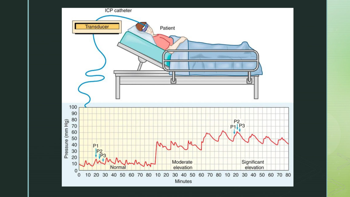
Reading the waveform

ICP is displayed as a mean pressure in mm Hg, and the waveform has three components.

WaveNameWhat it representsNormal height
P1Percussion waveArterial pulsations - she glossed it as "the direct impulse of arterial pulsations on the CSF"Highest of the three
P2Rebound or tidal waveIntracranial compliance - relative brain volume. Her gloss: "what the brain tissue is responding to from that initial surge - is it able to accommodate this blood volume change, or is that too much?"Middle
P3Dicrotic waveVenous pulsations; follows the dicrotic notchLowest of the three
Her ICP waveform slide, comparing a normal tracing with an abnormal one. On the normal tracing the three peaks step downward. On the abnormal one, P2 has climbed above P1 - the single finding to recognise here.

Inaccurate readings and infection

  • Causes of inaccurate readings: a CSF leak around the monitoring device; obstruction of the catheter by blood clot or tissue; a difference in height between the catheter and the transducer; kinks in the tubing; incorrect height of the drainage system relative to the patient's reference point; bubbles or air in the tubing.
  • Infection is the major complication of ICP monitoring. Contributing factors: monitoring for more than 5 days, use of a ventriculostomy, a CSF leak, and a concurrent systemic infection.
  • Nursing: routinely assess the insertion site, use aseptic technique, and monitor CSF drainage for changes in color and clarity.

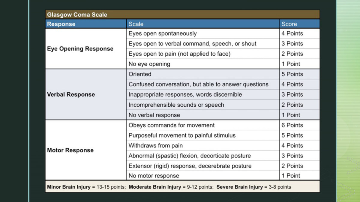
1.11 The Glasgow Coma Scale

The Glasgow Coma Scale assesses level of consciousness and, used serially, changing states. It scores three abilities: the ability to open the eyes to a verbal or painful stimulus, the ability to speak, and the ability to obey commands. The total is the sum of the three.

ScoreMeaning
15Normal score in an alert person - the maximum
8 or lessGenerally indicates coma. Also the threshold for intubation in head injury, and half of the indication for ICP monitoring
3The lowest possible score
The Glasgow Coma Scale as she presents it. The two abnormal motor responses are the postures from section 1.8: abnormal flexion scores 3 (decorticate), abnormal extension scores 2 (decerebrate). Score the patient's BEST response in each category, not the first one you see.
CategoryBest response score
Eye opening (max 4)Spontaneous 4 - to name or command 3 - to pain 2 - none 1
Best verbal response (max 5)Oriented and conversant - correctly identifies self, place, year and month 5 - confused but conversant 4 - inappropriate or disorganized words 3 - incomprehensible sounds or moaning 2 - no sound even to pain 1
Best motor response (max 6)Obeys commands 6 - localizes pain 5 - flexion withdrawal 4 - abnormal flexion, decorticate 3 - abnormal extension, decerebrate 2 - no response 1

1.12 CSF Drainage and Cerebral Oxygenation Monitoring

If the patient has a ventriculostomy, you can control ICP by removing CSF. The provider writes the level at which to start draining and the frequency - intermittent or continuous.

Intermittent drainage - her worked example

"The doctor has an order that says, when the intracranial pressure is higher than 20, you need to drain. So what you'll do - you'll go to the system, you'll move the stopcock to allow for drainage, and you'll let it drain for about 2 to 3 minutes, and then close the stopcock again to make it a closed system." She also fixed the vocabulary in place: "when it's opened up, it's an open system; when it's closed, it's a closed system."

  • Intermittent: the RN opens the system at the stopcock at the ordered ICP, drains for 2-3 minutes, then closes it again.
  • Continuous: the drain stays open and you carefully monitor the amount of CSF drained - the risk here is taking off too much.
  • Normal CSF production is 20-30 mL/hr, and total CSF volume is about 150 mL within the ventricles and subarachnoid space. Those two numbers are what make an hour of unwatched continuous drainage dangerous.
  • Post a sign at the bedside so that anyone turning, suctioning or repositioning the patient knows it will affect drainage.
  • The drain must be closed for 6 minutes before an ICP waveform reading is accurate. A number read off an open drain is not the patient's ICP.
  • The chapter adds the complications her slide does not name: ventricular collapse, infection, and herniation or subdural hematoma formation from rapid decompression.

Cerebral oxygenation monitoring

Two separate technologies, measuring two different things. Her slide lists them together and it is easy to blur them; keep them apart.

DeviceWhere it sitsWhat it measuresNumbers
LICOX and Neurovent cathetersIn healthy white matter of the brainBrain tissue oxygen pressure (PbtO2), continuously, plus brain temperaturePbtO2 normal 20-40 mm Hg. Low PbtO2 = ischemia or regional tissue hypoxia. A lower brain temperature may produce better outcomes
Jugular venous bulb oximetryIn the internal jugular vein, tip at the jugular bulb - a separate catheter, not a LICOX featureSjvO2 - jugular venous oxygen saturation, reflecting total venous brain tissue extraction of oxygen; a measure of cerebral oxygen supply versus demandNormal 60-75%. Below 50% indicates impaired cerebral oxygenation

1.13 Drug Therapy for Increased ICP

The goals of interprofessional care are to identify and treat the underlying cause - usually an increase in blood, brain tissue or CSF - and to support brain function by maintaining adequate oxygenation and preventing secondary injury. Early recognition and treatment produce better outcomes. Mechanical ventilation may be needed to keep PaCO2 in the normal range and maintain PaO2, and surgery may be needed if the cause is a mass.

DrugHow it lowers ICPMonitor
Mannitol (Osmitrol) 25% osmotic diuretic, IVTwo effects. It reduces blood viscosity and hematocrit, which increases CBF and cerebral oxygen delivery. And it creates a vascular osmotic gradient - "a concentration difference which pulls fluid back into the blood vessels from the surrounding tissues," as she put it - reducing brain fluid content and ICP. Mannitol is then filtered by the kidneys, so urine output rises.Fluid and electrolyte status. The chapter adds that it may be contraindicated in renal disease and with an already increased serum osmolality
Hypertonic saline commonly 3% sodium chlorideAllows massive movement of water out of edematous brain cells into the blood vessels, improving CBF. Her mechanism: "it's going to do this by osmosis - create a greater concentration of solutes on the outside of the cell membrane, and that's what's going to cause the water to move from the intracellular to extracellular spaces." Just as effective as mannitol and often used with it in severe brain injury.BP and serum sodium. The chapter adds the risk being watched for: intravascular fluid volume excess
CorticosteroidsTreat vasogenic edema around a tumor or abscess. She added two benefits not on the slide: they can also improve cerebral blood flow and restore autoregulation.Hyperglycemia, infection, GI bleeding. The chapter adds that they are not recommended in TBI, that glucose is checked at least every 6 hours, and that an H2 blocker or PPI is given alongside to prevent GI ulcers
IV 0.9% sodium chlorideThe carrier for secondary medications.The chapter gives the reason it is specified: with D5W or 0.45% NaCl, serum osmolality falls and cerebral edema may increase
DrugHow it lowers ICPMonitor
AntipyreticsFever raises cerebral metabolism, which raises demand for blood and oxygen, which raises ICP. Target temperature 36-37 °C.She named Tylenol as the example and gave the shivering mechanism - a febrile patient shivers, and shivering raises metabolic demand further
Antiseizure medicationsSeizures raise metabolic demand and ICP; prophylaxis is recommended in severe brain injury.Seizure activity; drug levels
Barbiturates pentobarbital, thiopentalUsed when increased ICP is refractory to other treatments. Her slide's phrase is that they "calm down" brain activity; she gave the actual mechanism: "it's going to reduce cerebral metabolism, which reduces the demand for oxygen and blood flow, thus lowering intracranial pressure."ICP, blood flow and EEG. The chapter adds that dosing is titrated to the bedside EEG, and that total burst suppression marks maximal therapeutic effect

1.14 Nursing Management of Increased ICP

Her four goals, in her order: (1) maintain a patent airway; (2) have ICP within normal limits; (3) have normal fluid, electrolyte and nutrition balance; (4) prevent complications from immobility and decreased level of consciousness.

Airway - the first goal for a reason

Maintaining the airway is a major nursing responsibility. As level of consciousness falls, the patient is at increased risk of obstruction. She gave the mechanism the slide implies: "their tongue can fall back and drop back into their throat, occluding their airway - and that's why a lot of these patients may need to be intubated or have some type of artificial airway." Increased secretions obstruct as well.

  • Snoring sounds indicate obstruction and require immediate intervention. Not a comfort issue - a patency issue.
  • Mechanical ventilation may be needed; monitor ABGs. The chapter adds the trigger: any patient with a GCS of 8 or less, or an altered LOC that prevents maintaining a patent airway or effective ventilation, needs intubation.
  • Suctioning: keep it minimal and under 10 seconds.
  • A nasogastric or orogastric tube prevents abdominal distention, which interferes with respiratory function - and, by the venous chain in section 1.1, raises ICP.
  • The chapter adds two suctioning refinements her slides omit: give 100% oxygen before and after, and limit to 2 passes per procedure to avoid cumulative ICP rises.

Sedation

Pain, anxiety, fear and noxious stimuli all raise ICP, so sedatives, analgesics and paralytics are used. The difficulty is that these drugs alter the neurologic state and can mask the very changes you are watching for - which is why the pharmacology is chosen for short duration of action.

AgentClassWhy this one
Fentanyl, morphine sulfateOpioidsPain management with rapid onset and minimal effect on CBF or oxygen metabolism
PropofolIV sedativeAnxiety and agitation. Rapid onset, short half-life, so an accurate neuro assessment can be done soon after the infusion is stopped
Dexmedetomidine (Precedex)α2-adrenergic agonistContinuous IV sedation of mechanically ventilated patients for up to 24 hours. Watch for hypotension - hypotension lowers MAP and therefore lowers CPP
VecuroniumNondepolarizing neuromuscular blocking agentA paralytic. The chapter adds the safety point: it paralyzes muscles without blocking pain, so it must be given with a sedative or analgesic
BenzodiazepinesSedativeUsually avoided - hypotensive effect and long half-life

Fluid and electrolyte balance

Monitor IV fluids closely, track intake and output and daily weights, and follow serum electrolytes - especially glucose, sodium, potassium, magnesium and osmolality. Urine output is the parameter that detects the two opposite endocrine complications of brain injury. She reframed the reason: you monitor output not to prevent these so much as "to identify an issue like this that could be occurring."

Diabetes insipidusSIADH
ADHDecreased ADHExcess secretion of ADH
Urine outputIncreasedDecreased
Serum sodiumHypernatremiaDilutional hyponatremia
ConsequenceDehydration - severe, and fast, if not identified and treatedCerebral edema, change in LOC, seizures, coma
TreatmentFluid replacement, vasopressin, or desmopressin acetate (DDAVP)Managed by treating the ADH excess and restricting fluid

Body position

  • Keep the patient head up. Elevating the head of the bed promotes drainage from the head and decreases the vascular congestion that produces cerebral edema.
  • Avoid extreme neck flexion - it obstructs venous outflow and raises ICP. Keep the head midline.
  • Turn with slow, gentle movements. Rapid position changes raise ICP.
  • Avoid pain and discomfort during turning, because pain raises ICP.
  • Avoid extreme hip flexion - it raises intraabdominal pressure, which raises ICP.
  • The chapter adds a limit her slides do not carry, and it is a good exam-level nuance: raising the head of the bed more than 30 degrees can decrease CPP by lowering systemic blood pressure. Higher is not automatically better; position for the best combination of ICP and CPP.
  • The chapter also notes that turning, skin care and even passive range of motion can elicit posturing reflexes in some patients, and that patients are still turned at least every 2 hours to prevent atelectasis and contractures.

Protection from injury

  • Agitation raises the risk of injury. Restraints may be needed, or a family member staying at the bedside. The chapter adds the caution that agitation may increase with restraints, which signals the need for a different approach.
  • Seizure precautions: padded side rails, an Ambu bag at the bedside, readily available suction, accurate and timely administration of antiseizure drugs, and close observation.
  • Antiseizure prophylaxis is recommended in severe brain injury. The chapter narrows this: prophylaxis is aimed at early seizures in the first 7 to 10 days, and is controversial for mild to moderate injury.
  • She made the equipment concrete: "you might have seen in hospitals where they have those paddings on the side of the bed - so if a patient does have a seizure, they have something protecting them. And you want to have an Ambu bag at the bedside in case their respiratory function is declined."

Neurologic assessment techniques she demonstrated

TestHow it is doneNormal / abnormal
Oculocephalic reflex (doll's eyes)Turn the patient's head briskly left or right while holding the eyelids open. Then quickly flex and extend the neck.Normal: the eyes move across the midline in the direction opposite the turn - up when the neck is flexed, down when extended. The chapter adds that this is usually reserved for brain death evaluation
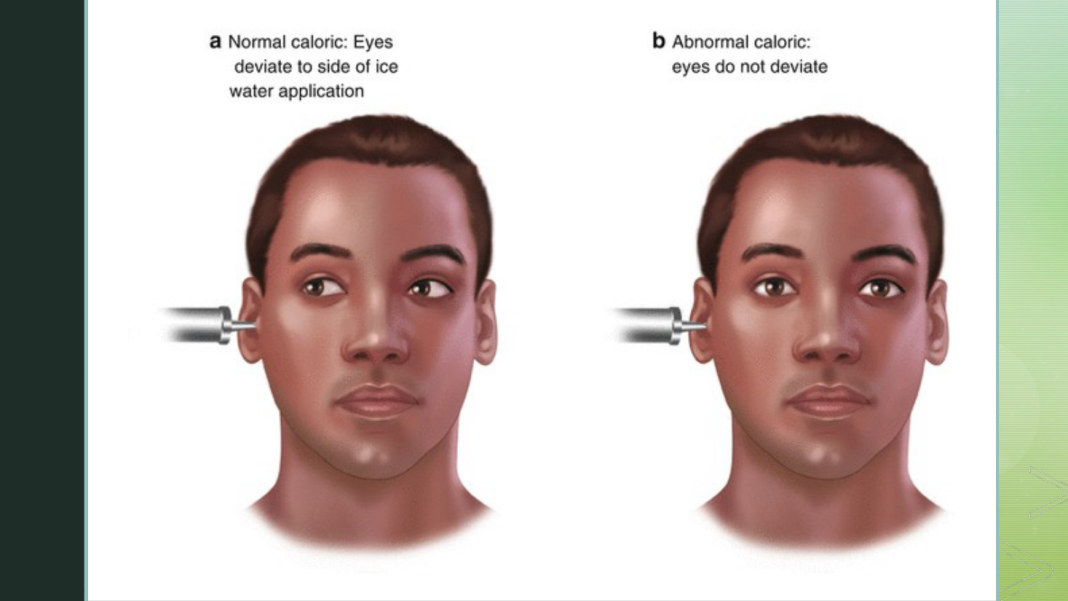
Oculovestibular reflex (cold calorics)With the head of the bed elevated, instill a syringe of ice-cold water into the external auditory canal and watch the eyes for a full minute.Absence of eye movement indicates severe neurologic demise. She gave both the intuition - "if somebody shot cold water into your ear, you would definitely respond" - and the expected direction: the eyes deviate toward the side the water was instilled
Extremity strengthAsk the patient to squeeze your fingers on both sides; have them raise their arms and hold against your downward pressure. Test all four extremities and note asymmetry.Only valid in a patient who is conscious and able to follow commands - she said so explicitly. In the unconscious patient, observe spontaneous movement, then apply a painful stimulus

1.15 Head Injury: Scalp Lacerations and Skull Fractures

She marked the transition plainly - "okay, so that was intracranial pressure" - and moved to head injury with about forty-four minutes left. Head injury means any injury to the scalp, skull, or brain. Traumatic brain injury (TBI) is the serious form. The most common causes are falls and motor vehicle accidents; others are firearms, assault, sports-related trauma, recreational injuries, and war-related injuries. The potential for poor outcomes is high.

Scalp lacerations

The easily recognized form of external head trauma. The scalp has many blood vessels with poor constrictive abilities, so profuse bleeding occurs even with relatively small lacerations. The complications are blood loss and infection.

Skull fractures

Skull fractures are described three ways: (1) linear or depressed; (2) simple, comminuted, or compound; (3) closed or open. Location determines the manifestations - the fracture line itself is less informative than what sits under it. The major complications are intracranial infection, hematoma, and meningeal and brain tissue damage.

TypeDescriptionTypical cause
LinearA break in the continuity of bone without a change in the relationship of the partsLow-velocity injury
DepressedInward indentation of the skullA powerful blow
SimpleA linear or depressed fracture without fragmentation or communicating lacerationsLow to moderate impact
ComminutedMultiple linear fractures with fragmentation of bone into many piecesDirect, high-momentum impact
CompoundA depressed fracture plus a scalp laceration with a communicating pathway into the intracranial cavitySevere head injury

Manifestations - reading the location off the face

Her slide names the classic findings: Battle sign, raccoon eyes, CSF rhinorrhea, tinnitus, deafness, loss of taste and visual changes. She located and defined them: Battle sign is "bruising behind the ear," raccoon eyes is "bruising around the eyes," and tinnitus is "ringing in the ears."
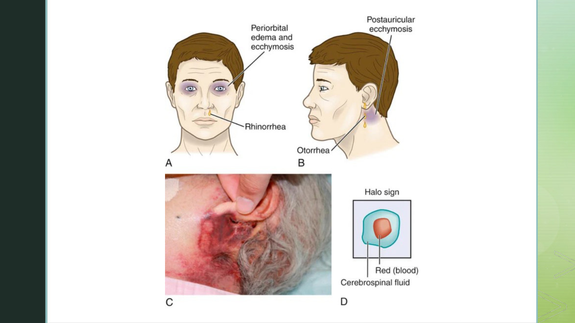
Battle's sign and raccoon eyes. Both are bruising in a characteristic distribution rather than at the point of impact, which is what makes them useful - they point to a basilar skull fracture underneath. The chapter notes these findings evolve over several hours, so their absence on arrival proves nothing.
Fracture locationManifestations (from the chapter's table)
BasilarCSF otorrhea, bulging tympanic membrane from blood or CSF, Battle sign, tinnitus or hearing difficulty, rhinorrhea, facial paralysis, vertigo
FrontalExposure of brain to contaminants through the frontal air sinus, air in forehead tissue, CSF rhinorrhea, pneumocranium
OrbitalPeriorbital bruising (raccoon eyes), optic nerve injury
ParietalDeafness, CSF or brain otorrhea, facial paralysis, loss of taste, Battle sign
TemporalBoggy temporal muscle, oval bruise behind the ear in the mastoid region, CSF otorrhea, middle meningeal artery disruption epidural hematoma
Posterior fossaOccipital bruising causing cortical blindness, visual field defects, rarely ataxia or cerebellar signs

This location table is Lewis's, not hers - it is the Table 61.6 material she pointed students to. The one row worth learning cold is temporal, because it connects the fracture to the artery to the hematoma: a temporal fracture tears the middle meningeal artery, and that is how an epidural hematoma is made.

The CSF leak

Rhinorrhea is CSF leaking from the nose. Otorrhea is CSF leaking from the ear. Either one means the fracture has torn through the dura mater, which is why the leak matters far more than the volume of fluid suggests: there is now an open path between the outside world and the CSF.

1.16 Diffuse and Focal Brain Injury

The organizing split for brain trauma is whether the damage is spread out or localized.

Diffuse (generalized)Focal (localized)
DefinitionDamage to the brain is not localized to one areaDamage is localized to one specific area
Examples on her slideConcussion - axonal injuryContusion - hematoma
Typical mechanismRotational or acceleration-deceleration forces acting on the whole brainA blow or a bleed at one place

Concussion

A minor diffuse head injury - a sudden, transient mechanical head injury with disruption of neural activity and a change in level of consciousness. The classic signs are a brief disruption in LOC, retrograde amnesia about the event, and headache. It resolves spontaneously and is usually considered benign.

Postconcussion syndrome develops 2 weeks to 2 months after the injury in some patients: persistent headache, lethargy, personality or behavior changes, a shortened attention span, decreased short-term memory, and changes in intellectual ability.

Diffuse axonal injury

Her slide lists "axonal injury" as two words. She defined it and gave two mechanisms: "axonal injury is when the brain's nerve fibres tear - the brain can shift or rotate in the skull. So this can happen with a car crash, or like shaken baby syndrome."

The chapter fills in what her slide and lecture leave out, and it contains the timing fact most likely to be tested: axonal damage is not an immediate tearing. Trauma changes the function of the axon, which then swells and disconnects, and that process takes 12 to 24 hours to develop and may persist longer. Clinically it produces decreased LOC, increased ICP, decortication or decerebration, and global cerebral edema. Severe DAI carries a poor functional prognosis, and survivors go to the ICU to be watched for rising ICP.

Lacerations of brain tissue

A focal injury involving actual tearing of brain tissue, occurring with depressed and open fractures and penetrating injuries. Management is antibiotics until meningitis is ruled out and preventing secondary injury from increased ICP. If bleeding is deep within brain tissue, both focal and generalized signs develop. The chapter adds the blunt reason there is no surgical option: surgical repair of a brain laceration is impossible because of the nature of brain tissue.

In major head trauma, delayed responses occur - bleeding, hematoma formation, seizures, cerebral edema. Subarachnoid and intraventricular hemorrhage can both result from head trauma. Intracerebral hemorrhage is generally due to a cerebral laceration and behaves as a space-occupying lesion: unconsciousness, hemiplegia on the contralateral side, and a dilated pupil on the ipsilateral side - the localizing pair from section 1.7.

Contusion

A bruising of brain tissue within a focal area, associated with closed head injury and often occurring at a fracture site. There may be areas of bleeding, infarction, necrosis and edema. Overall prognosis is based on the amount of bleeding around the contusion site.

Contusions may continue to bleed or rebleed. The chapter's word for this is that they "blossom" on subsequent CT scans, and that bleeding worsens the neurologic outcome. Neurologic assessment may show focal or generalized manifestations depending on size and location. Seizures occur especially with frontal or temporal lobe involvement. And anticoagulant use and coagulopathy are associated with increased bleeding, more severe injury, and increased mortality - her slide flags this as very important in older adults.

Emergency management and diagnostics

  • If unresponsive, assess circulation, airway, breathing. If responsive, monitor airway, breathing and circulation.
  • Assume a neck injury with every head injury - stabilize the cervical spine.
  • Apply oxygen by nonrebreather if needed.
  • Establish IV access with two large-bore catheters. The chapter specifies the fluid: normal saline or lactated Ringer's.
  • Intubate if the GCS is below 8.
  • Control external bleeding with a sterile pressure dressing.
  • CT scan is the primary study. MRI is more sensitive than CT for small lesions. Transcranial Doppler measures CBF velocity - she added that it "shows the speed and direction of blood flow, and any presence of blood clots as well."
  • The chapter adds a caution for ongoing care: give fluids cautiously, because fluid overload raises ICP.

1.17 The Three Hematomas

She defined the word first, because the slide assumes it: "a hematoma is pretty much like a large bruise underneath the skin, or a collection of blood underneath the skin - and in this case, in the brain." The three are named for where the blood collects relative to the dura.

EpiduralSubduralIntracerebral
Where the blood isBetween the dura and the inner surface of the skull - the epidural spaceBetween the dura mater and the arachnoid layer of the meningesWithin brain tissue itself
SourceUsually a linear fracture crossing a major artery in the dura, causing a tear. Venous or arterial in originInjury to brain tissue and its blood vessels. Usually venous; the chapter adds that most arise from bridging veins draining into the sagittal sinusRupture of intracerebral vessels at the time of injury
SpeedFast if arterial. A neurologic emergencySlower because it is venous; an arterial subdural develops more rapidlyDepends on the bleed
LocationAnywhere a dural artery is torn - classically temporalOver the surface of the brainUsually frontal and temporal lobes
Classic presentationInitial unconsciousness at the scene, a brief LUCID INTERVAL, then a decrease in LOC. Plus headache, nausea, vomiting, focal findingsAcute, subacute or chronic - see the table belowSize and location determine the outcome
TreatmentRapid surgical evacuation to prevent cerebral herniation, plus management of increased ICPEvacuation and decompression; craniotomy for the acute formManagement of increased ICP; surgical evacuation depending on size and site

Subdural hematoma by timing

TypeOccurrence after injuryProgressionTreatment
Acute24-48 hr after severe traumaImmediate deteriorationCraniotomy, evacuation and decompression
Subacute48 hr - 2 weeks after severe traumaDecline in mental status as the hematoma develops; progression depends on size and locationEvacuation and decompression
ChronicWeeks to months - usually more than 20 days. The injury often seemed trivial or was forgotten by the patientNonspecific, nonlocalizing; progressive change in LOCEvacuation and decompression, plus membranectomy
  • Acute subdural manifestations: decreasing LOC, headache, drowsiness, confusion, unconsciousness, and ipsilateral pupil dilation and fixation if the ICP is significantly increased.
  • Chronic subdural manifestations: the presenting problem is focal symptoms rather than signs of increased ICP. The chapter gives the reason: brain atrophy in older adults creates a larger subdural space, so a hematoma has room to grow without pushing pressure up - and the same atrophy puts bridging veins under tension, making them easier to tear.
  • The chapter adds that a history of alcohol use raises the risk, because of falls.

1.18 Cranial Surgery

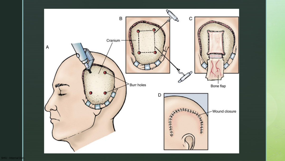
She deliberately front-loaded burr holes so that the next slide would make sense - "I want to talk about [burr holes] before we talk about this, because burr holes are included in these procedures." That is the right order to learn them in, because a burr hole is the building block of everything else.

ProcedureWhat is doneIs the bone put back?
Burr holeAn opening into the cranium made with a drill. Used to remove localized fluid and blood beneath the dura. The chapter adds that burr holes may be used alone in an extreme emergency for rapid decompression, followed later by a craniotomyNot applicable
CraniotomyAn opening into the cranium with removal of a bone flap and opening of the dura - to remove a lesion, repair a damaged area, drain blood, or relieve elevated ICP. The surgeon drills burr holes and uses a saw to connect them to free the flapYES - the flap is secured with small plates or wired shut at the end of the operation
CraniectomyExcision into the cranium to cut away a bone flap. Done when extreme swelling is expected - removing skull gives the brain somewhere to expand and reduces the risk of herniationNO - not yet. The flap is left off until the swelling resolves
CranioplastyRepair of a cranial defect from trauma, malformation or previous surgery. Artificial material replaces damaged or lost boneIt is the repair itself - the later step after a craniectomy
Stereotactic procedurePrecise localization of a specific brain area using a frame or frameless system based on three-dimensional coordinates. Used for biopsy, radiosurgery or dissectionEntry through a burr hole or a bone flap
Her cranial surgery slide comparing craniectomy, craniotomy and cranioplasty. The whole distinction is in the third column of the table above: does the bone go back on today, or not?

Stereotactic procedures

A stereotactic biopsy obtains tissue samples for histologic examination. The surgeon drills a burr hole or creates a bone flap for the entry site, then introduces a probe and a biopsy needle. She gave the indication in patient terms: "if a patient has a tumor, or something that needs to be tested to see if these are benign or malignant cells." Other stereotactic procedures remove small brain tumors and abscesses, drain hematomas, perform ablative procedures for diseases such as Parkinson's, and repair arteriovenous malformations. The major advantage is reduced damage to surrounding tissue.

Stereotactic radiosurgery targets tumor cells and other abnormal growths. She made the point the slide does not say outright - it is radiation, not surgery: "the stereotactic radiosurgery is just going to be like a radiation procedure to target those tumor cells, if a patient does have a malignant brain tumor." The chapter agrees and adds the hardware: a linear accelerator, Gamma Knife or CyberKnife, with the head held still in a stereotactic frame, delivered in one session of a few hours or in several sessions.

Ongoing nursing management after head injury

  • Monitor for increased ICP - every head-injury patient is at risk.
  • Assess the GCS, neurologic status, and whether a CSF leak has occurred.
  • Use a calm, gentle approach. She gave the reasoning: "they might be scared - what's going on? And if they have increased intracranial pressure, you also don't want to increase that even more by putting them in a stressful environment."
  • Report any change in neurologic state, even if subtle.
  • Her goals for the patient with acute head injury: maintain adequate cerebral oxygenation and perfusion; stay afebrile; be free of discomfort; be free from infection; have adequate nutrition; attain maximal cognitive, motor and sensory function.
UNIT 2

Acute Kidney Injury and Chronic Kidney Disease

Covers: Her 86-slide "Kidneydiesease-2" deck and the 136-minute AKI - CKD recording - Lewis's Ch. 51 (pp. 1232-1265) and Ch. 17 (pp. 313-315) for gaps only

She delivers this deck in a single uninterrupted pass, alone, working straight down her own slides. There is no reordering, and there are only two slides she does not speak to. The deck falls into four blocks and this unit follows them in her order: AKI on slides 1-29, CKD on slides 30-54, dialysis on slides 55-77, kidney transplant on slides 78-86. Sections 2.1 to 2.9 are AKI, 2.10 to 2.16 are CKD, 2.17 to 2.19 are dialysis, 2.20 is transplant.

Roughly a third of what she says is printed nowhere on the slides - definitions of BUN, creatinine, GFR, specific gravity, anuria, peritonitis and Dacron cuffs; a pre-CT contrast rule that appears on no slide in the deck; the populations in whom an AV fistula is hard to achieve; the limb-protection protocol for a dialysis access arm. All of that is carried here, and where a fact comes from Lewis rather than from her, the sentence says so.

2.1 How the Kidneys Work, and What Acute Kidney Injury Is

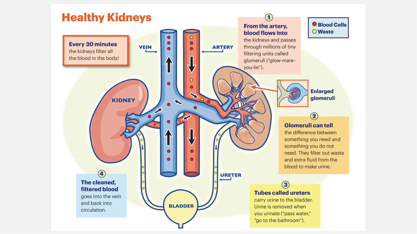
She opens with normal physiology and says why: "it's always helpful with, uh, you know, when you're doing any kind of system or any issues with a system to go back and just look at how the system should be working." Her slide is a six-box flow diagram, and the boxes are the spine of what follows - each of the three causes of AKI is a failure at one of these steps.

Blood enters through the renal arteries, which branch into arterioles; arterioles reach a nephron and form the glomerulus, whose fenestrations let certain products through and keep others out. Blood is filtered at the glomeruli, tubular reabsorption puts some of the filtrate back into the blood, and what is left becomes urine, carried down the ureters to the bladder and out through the urethra. Cleaned blood leaves by the renal vein.

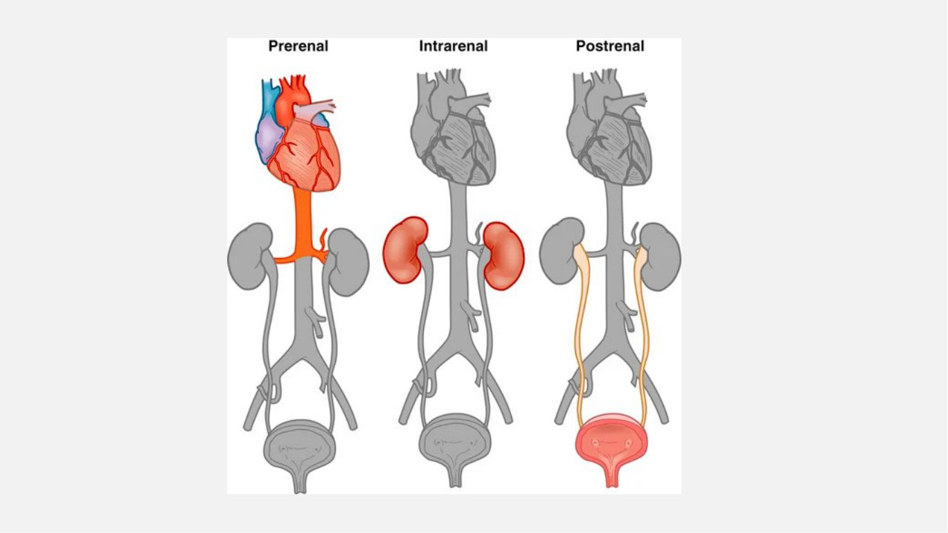
Her simplified kidney and nephron picture - "just a simplified version of what they do." Read it as the map for section 2.2: a prerenal problem sits upstream of the glomerulus, an intrarenal problem inside the tissue drawn here, a postrenal problem downstream of the collecting system.

Acute kidney injury

AKI is a rapid loss of kidney function, with or without decreased urine output. Note the second half of that sentence - output can be normal and the kidneys can still be failing. It is accompanied by rising blood urea nitrogen (BUN), creatinine (Cr) and potassium, and severity runs from a small rise in creatinine to frank azotemia, the accumulation of nitrogenous waste products in the blood. AKI develops over hours to days and is potentially reversible. Its causes are prerenal, intrarenal and postrenal.

Her slide prints BUN at 6-24 mg/dL and creatinine at 0.7-1.3 mg/dL (males), 0.6-1.1 mg/dL (females), with no explanation. She defined both analytes from scratch: urea is "a waste product that your body creates, uh, when it breaks down protein," and creatinine is "a waste product that comes from breakdown of muscle tissue and protein in food." Those two definitions are why creatinine turns out to be the better marker in section 2.6.

2.2 Causes of AKI: Prerenal and Intrarenal

Prerenal

Prerenal causes reduce systemic circulation, so renal blood flow falls, and less flow means less glomerular perfusion and less filtration. Nothing is wrong with the kidney tissue; the kidney is being starved from upstream. Autoregulation fires to protect essential organs, and she walked the chain where the slide only lists hormones: angiotensin II "increases blood pressure and it stimulates the release of aldosterone, and then aldosterone increases sodium and water retention," with ADH and norepinephrine doing the same work by other routes. The result is prerenal azotemia - decreased sodium excretion, increased sodium and water retention, decreased urine output.

Compensation does not last. She generalised the failure point herself: "like like we always say, with any, um, uh, regulatory mechanism or compensatory mechanism, it eventually does fail." When decreased perfusion persists, the compensatory mechanisms give out and the prerenal problem becomes an intrarenal one. A prerenal cause left alone long enough stops being prerenal.

Intrarenal

Intrarenal causes damage kidney tissue directly, impairing nephron function. Her four sources are prolonged ischemia, nephrotoxins, hemoglobin from hemolysed red cells and myoglobin from necrotic muscle. She stopped on the nephron image to call it "one of the most important areas of the kidney" - one of only two things in the lecture she called most important - and supplied the mechanism the slide omits: nephrotoxins damage tubular epithelial cells or crystallise and obstruct intrarenal structures, and the obstruction provokes inflammation. Myoglobin behaves the same way, since "high levels of myoglobin can precipitate in the renal tubules, also causing more obstruction and direct kidney damage." Lewis adds that both pigments also cause renal vasoconstriction.

Acute tubular necrosis

ATN is the most common intrarenal cause of AKI in hospitalised patients. Severe ischemia disrupts the basement membrane and destroys tubular epithelium in patches; nephrotoxins necrose tubular epithelial cells, which slough off and plug the tubules. It follows ischemia, nephrotoxins or sepsis, and ischemic and nephrotoxic causes account for 90% of intrarenal AKI. Her other in-hospital risk factors are major surgery, shock, blood transfusion reaction, muscle injury from trauma, and prolonged hypotension. ATN is potentially reversible if the basement membrane is not destroyed and the tubular epithelium regenerates - one conditional, and it is why that structure is worth knowing by name. Note that Lewis states the claim two ways: Table 51.1 (p.1233) gives the most common cause of AKI, full stop, as ATN, while the body text (p.1234) scopes it to the most common intrarenal cause in hospitalised patients. Her slide follows the narrow version and so does this guide, because it survives a question that also offers a prerenal option.

2.3 Causes of AKI: Postrenal

This was her third-longest stop - 467 words, about 2.9 minutes - on the category Lewis says causes fewer than one AKI case in ten, and she spent it on mechanism rather than on the list. Postrenal causes involve mechanical obstruction to the outflow of urine. Urine refluxes into the renal pelvis, pressure inside the kidney rises, kidney structures are damaged, and filtration falls. Her common causes are BPH, prostate cancer, stones, trauma and external tumors. She self-corrected the anatomy mid-sentence here - obstruction "is going to prevent, uh, urine flow of down the urethra. Uh, sorry. The ureters. Ureters, as it should be" - and she had been pointing at the next slide's figure since two slides earlier, promising "I have a picture which I'll show you after, actually." Bilateral ureteral obstruction produces hydronephrosis - dilation of the kidney - with raised hydrostatic pressure and tubular blockage behind it.

Her three-panel comparison of the cause categories. The coloured zone in each panel is where the problem sits - above the kidney, inside the kidney tissue, below the kidney. The middle panel is the only one in which the nephron itself is shaded, which is the whole difference between a kidney being starved or blocked and a kidney being damaged.

2.4 The Clinical Course of AKI and the RIFLE Classification

Prerenal and postrenal AKI usually resolve quickly with treatment, provided they have not already caused intrarenal damage. Once intrarenal damage occurs, AKI runs a prolonged course through three phases - oliguric, diuretic, recovery. Lewis adds the outcome she leaves implicit: when a patient does not recover from AKI, CKD may develop.

RIFLE is the classification her deck and Lewis both use to stage AKI, and it is the only one either contains. There is no KDIGO staging in this deck, in this lecture, or in the AKI half of Ch. 51 - a search returns nothing in any of the three. If you have met KDIGO stages 1-3 elsewhere, they are not what this material uses. The letters are three stages of severity - Risk, Injury, Failure - followed by two outcome variables, Loss and

End-stage renal disease. Each of the first three is met by either the creatinine criterion or the GFR criterion or the urine output criterion. Read the table as a pattern rather than a list of numbers: the creatinine multiples run 1.5, 2, 3 and the GFR losses run 25%, 50%, 75%, moving together at every stage; the urine output threshold falls only once, at Failure, while what changes at Risk and Injury is the duration - 6, then 12, then 24 hours. Loss and ESRD are measured not in creatinine but in time.

StageCreatinine or GFR criterionUrine output criterion
R - RiskSerum Cr increased x 1.5 or GFR decreased by 25%<0.5 mL/kg/hr for 6 hr
I - InjurySerum Cr increased x 2 or GFR decreased by 50%<0.5 mL/kg/hr for 12 hr
F - FailureSerum Cr increased x 3 or GFR decreased by 75% or serum Cr >4 mg/dL with an acute rise of at least 0.5 mg/dL<0.3 mL/kg/hr for 24 hr or anuria for 12 hr
L - LossPersistent acute kidney failure; complete loss of kidney function >4 weeks-
E - End-stage renal diseaseComplete loss of kidney function >3 months-

2.5 The Oliguric Phase: Urine, Fluid Volume, Acid-Base, and Sodium

Slide 13 is the longest single stop in the lecture - 745 words, about 4.7 minutes, 2.9 times her average - and roughly a third of it is off-slide. She interrupted herself before starting the phase at all: "Oh, and here, let me just clarify a couple definitions."

Urinary changes

Oliguria is a urine output of less than 400 mL in 24 hours. It usually appears within 1 to 7 days of the injury, and within 24 hours if the cause is ischemia. The phase lasts 10 to 14 days on average and can run months, and the longer it lasts the poorer the prognosis for complete recovery. The pattern points at the cause: oliguria usually accompanies prerenal causes, anuria usually accompanies postrenal obstruction, and nonoliguric AKI - an output above 400 mL/24 hr, in about 50% of patients - accompanies the intrarenal causes, acute interstitial nephritis and ATN.

Urinalysis may show casts, RBCs and WBCs - Lewis explains what the casts are, where her slide only names them: mucoprotein impressions of necrotic tubular epithelial cells sloughed into the tubules. Specific gravity fixes at around 1.010 and urine osmolality at about 300 mOsm/kg, both of which are the values of plasma. Proteinuria may be present if the AKI involves glomerular membrane dysfunction, and her reasoning generalises to every proteinuria question in this unit: "proteins are large molecules and they should not be making it through that filtration process."

Fluid volume, acid-base and sodium

Hypovolemia can worsen all forms of AKI. Falling urine output produces fluid retention, and severity tracks the amount held: distended neck veins with a bounding pulse, edema and hypertension first, then heart failure, pulmonary edema, and pericardial and pleural effusions.

The damaged kidney cannot excrete hydrogen ions, so acid accumulates. She explained the bicarbonate half twice over, because two things happen to it at once: production falls, since reabsorption and regeneration of HCO3 are defective, and the existing supply is spent - "we're using the one that's already the bicarb, that's already present" - buffering the acid already there. Severe acidosis produces rapid, deep respirations to blow off CO2.

Damaged tubules cannot conserve sodium, so urinary sodium excretion rises and serum sodium ends up normal or low. Excess sodium intake is avoided because it drives volume expansion, hypertension and heart failure - and she gave the mechanism the slide leaves out, "when there's sodium, uh, there's also going to be water that's following it." Uncontrolled hyponatremia may lead to cerebral edema, because low serum sodium moves water into brain cells and swells them.

2.6 The Oliguric Phase: Potassium, and the ECG Changes She Never Named

Potassium is the most repeated topic in the recording: the word appears 53 times and "hyperkalemia" 27 times across 12 different slides, the only subject she returns to on more than ten. Two of her nine "very important" flags land eight lines apart on this one slide.

The kidneys normally excrete 80% to 90% of the body's potassium. In AKI they cannot secrete it, so serum potassium rises, and her slide adds three further sources - each a different route to the same number.

  • Trauma. Damaged cells release potassium as they break open, on top of what the kidney is failing to clear.
  • Bleeding and blood transfusions. Cellular destruction releases potassium into the extracellular fluid.
  • Metabolic acidosis. Hydrogen ions move into cells and potassium is driven out into the ECF - and she gave the reason the slide does not, "because both potassium and hydrogen are positively charged."

What hyperkalemia does to the heart

Her slide says hyperkalemia is often asymptomatic, that the patient is weak with severe hyperkalemia, that there are ECG changes, and that cardiac muscle is intolerant of acute rises in potassium, so emergency treatment is needed - a bullet she did not read. What she gave instead is a mechanism on no slide: high potassium disrupts "the electrical stability of the heart by, um, altering the resting membrane potential. This leads to impaired conduction, decreased excitability," and from there "potentially leading to life threatening arrhythmias or cardiac arrest."

The morphology comes from Lewis's Ch. 17, the assigned re-read, which sequences it rather than listing it (p.314): "The initial finding is tall, peaked T waves. As potassium increases, cardiac depolarization decreases. This leads to loss of P waves, a prolonged PR interval, ST segment depression, and widening QRS complex. Life-threatening dysrhythmias may occur." Read in that order, the three items on her slide are the first, fourth and fifth events in one progression, and the two she does not print - loss of P waves and a prolonged PR interval - fall between them.

Hematologic, waste and neurologic problems

Leukocytosis occurs, and her slide states flatly that the most common cause of death in AKI is infection - though eleven slides later she pulled back mid-sentence on it, "infections are the leading cause of, uh, death in patients with, um. Oh, one of the leading complications." Her slide and Lewis's Table 51.1 both say cause of death. BUN and creatinine both rise, but creatinine is the best serum indicator in AKI - and she gave the reason, which is not on the slide: BUN "can be affected by other things such as dehydration, steroids, um, severe injury or GI bleeding." Creatinine is better because it is not moved by those things. Neurologic changes appear as nitrogenous wastes accumulate in the brain and nervous tissue, escalating from mild to seizures and coma.

2.7 The Diuretic Phase, the Recovery Phase, and Diagnosing AKI

In the diuretic phase daily urine output is usually 1 to 3 litres and can reach 5 or more, and the phase lasts 1 to 3 weeks. The trap is that the volume looks like recovery and is not: the nephrons are still not fully functional. What has returned is the ability to excrete wastes; what has not is the ability to concentrate urine. On top of that, the high BUN in the filtrate causes osmotic diuresis, dragging water out with it.

The recovery phase begins when GFR increases, letting BUN and creatinine fall. She also defined GFR here, in passing, for a term the deck uses from slide 12 onward and never explains - it "pretty much tells you how much blood is being filtered out from the kidneys within a, uh, within a minute." Major improvements occur in the first 1 to 2 weeks, but it may take 12 months for kidney function to stabilise. Some patients never recover and progress to end-stage renal disease; Lewis adds that those who do recover may reach clinically normal function and still sit in an early stage of CKD.

Diagnostic studies

The history is essential for identifying which cause is operating, and Lewis makes the mapping explicit where her slide only says "history is essential": suspect prerenal with dehydration, hypotension or blood loss; intrarenal after nephrotoxic drugs or contrast; postrenal with changes in the urinary stream, stones, BPH, or bladder or prostate cancer. Urine output and serum creatinine occur late and are lagging indicators: a rise in creatinine may not appear until more than 50% of kidney function is lost, and the rate of rise determines severity, not the single value. Urinalysis showing casts, protein and abundant cells indicates intrarenal AKI, as do hematuria, pyuria and crystals.

  • Renal biopsy is the best way to confirm an intrarenal cause - confirmatory, not first-line.
  • Kidney ultrasound is often the first test done, because it images without exposing the patient to nephrotoxic contrast. It assesses kidney blood flow, tubular function and the collecting system, and evaluates obstruction.
  • CT shows lesions, masses, obstruction and vascular abnormalities; she added that non-contrast CT is acceptable for stones in a patient who already has AKI.
  • MRI or MRA with gadolinium contrast can be potentially fatal and is not advised.

2.8 Managing AKI: Interprofessional Care, Nursing Care, and Prevention

The goals are to eliminate the cause, manage the signs and symptoms, and prevent complications while the kidneys recover. Nothing here repairs a nephron; all of it buys time.

Fluid restriction and diuretics

Fluid intake is restricted in the oliguric phase, and her slide gives the arithmetic: add all losses for the previous 24 hours - urine, diarrhea, emesis, blood - and add 600 mL for insensible losses from respiration and diaphoresis. That total is the next day's allowance - and she spent much of this 480-word slide not on the formula but on where its numbers come from, "charted in your computer system... So the previous nurse should have been, uh, should be charting and then you should be charting." The calculation is only as good as the previous shift's intake and output record. Loop diuretics (furosemide) and osmotic diuretics (mannitol) are then used to manage overload, with the goal of increasing urine output and maintaining a nonoliguric state, which may help protect the kidneys. Her slide states the double-edge in the same breath: diuretics can also cause further fluid depletion and decrease renal blood flow, worsening the AKI.

Treating hyperkalemia

Hyperkalemia is one of the most serious complications of AKI and has to be treated. Slide 23 was her second-longest stop at 552 words. Her four measures do three different jobs, and she was sharper about the distinction than the slide is.

MeasureWhat it doesHer clarification
Insulin and sodium bicarbonatePromote a transient shift of potassium into cells; it eventually diffuses back into the bloodstream"A temporary fix of the problem" - the potassium has been hidden, not removed
Calcium gluconateRaises the threshold at which dysrhythmias occur; temporarily stabilises the myocardium"Calcium gluconate is not going to decrease the potassium at all. Uh, it has no role in that, but it does protect the heart from the side effects of hyperkalemia."
Sodium polystyrene sulfonate (Kayexalate)Removes potassium from the body"The potassium binds to, uh, the lumen of the GI tract" - and tell the patient to expect increased bowel movements
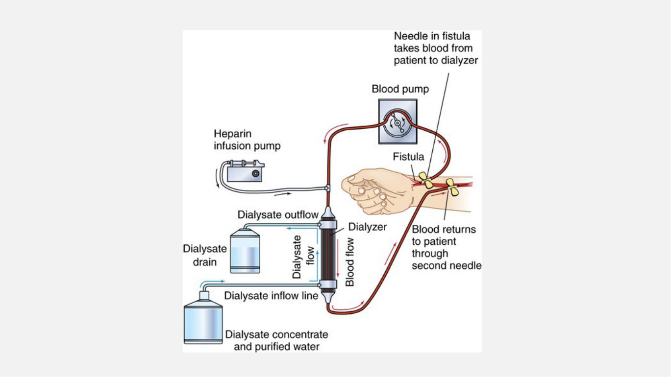
DialysisRemoves potassium from the bodySee sections 2.17 to 2.19

Renal replacement therapy and nutrition

Conservative therapy may be all that is needed until kidney function improves. When it is not, her indications for RRT are volume overload compromising cardiac or pulmonary status, high serum potassium, metabolic acidosis, BUN over 120 mg/dL, significant mental status change, pericarditis, pericardial effusion, or cardiac tamponade - and she sharpened that mental status item to "a very severe change." The modalities are HD, PD and CRRT. On nutrition: adequate calories to prevent catabolism and adequate protein, energy mainly from carbohydrate and fat to prevent ketosis, sodium restricted, and enteral nutrition preferred when oral intake fails - with the numbers her slide omits supplied by Lewis, 30 to 35 kcal/kg and 0.8 to 1.0 g protein/kg of desired body weight.

Nursing assessment

Daily weights, with the rationale she added - "checking at the same time each day on the same scale to get the most accuracy" - and 1 kg equals 1000 mL of fluid. Strict intake and output, vital signs, and urine assessment for colour, specific gravity, glucose, protein, blood and sediment. Edema, neck vein distention, bruises; the dialysis access site for inflammation or exudate; mental status; lungs for crackles, wheezes or decreased breath sounds; ECG monitoring. Lewis adds the oral mucosa, and listening for an S3 gallop, murmur or pericardial friction rub. Her clinical problems for AKI are electrolyte and fluid imbalance, risk for infection, and anxiety.

Prevention

Prevention and recognition of AKI are the most important aspects of care, and the centrepiece is contrast-induced nephropathy (CIN) - nephrotoxic injury from contrast media given for diagnostic studies.

  • Ensure adequate fluid intake if contrast is given to a high-risk patient - she added the order that accompanies it, "most of the times when a CT with contrast is ordered, I.V. fluids are ordered alongside with it." Lewis adds that the best way to avoid CIN is to avoid contrast, using ultrasound instead.
  • Hold metformin for 48 hours before and after contrast to reduce the risk of lactic acidosis, and monitor kidney function in anyone on potentially nephrotoxic drugs, given in the smallest effective dose for the shortest period (Lewis).
  • Caution patients about OTC drugs that worsen kidney function, especially NSAIDs - she named "ibuprofen, Aleve."
  • ACE inhibitors can decrease perfusion pressure and cause hyperkalemia, and may have to be stopped if it cannot be controlled - yet they are also used to prevent proteinuria and slow progression, especially in diabetes. She named a dose the deck does not, "a really small dose like 2.5 mg of lisinopril," and said the logic is hard: "it's kind of an interesting and kind of confusing at the same time."

Infection, teaching and older adults

Use aseptic technique. Patients with AKI have a blunted febrile response, so they may not have a fever - look for local signs and leukocytosis rather than waiting on a temperature. Antibiotic type, dose and frequency need careful consideration, because the kidneys are the main route of excretion for many antibiotics. Teach the signs of recurrent kidney disease and provide psychosocial care. Older adults are at increased risk because GFR declines with age; Lewis adds that the aging kidney compensates poorly for changes in fluid volume, solute load and cardiac output, and that patients over 65 are less likely to recover - though age is not a barrier to offering RRT, a point she makes herself on slide 56.

2.9 Her Two AKI Application Slides, Worked from the Chapter

Slides 28 and 29 close the AKI half with application questions. She spoke to neither - not a word on either slide - going straight from "So to be aware with that with elderly patients" into "Okay, so that was acute kidney disease." Her disposition for the whole deck was her closing line: "All right, that's it. So I'll go ahead and upload this and then we'll go over it in class." Her answers are therefore not on the recording at all. What follows is worked from Lewis's Ch. 51, and none of it should be read as her reasoning.

Slide 28 - "What is the cause of AKI in a patient who..."

Her patientCategoryWhy, from Ch. 51
Has an obstructing kidney stonePostrenalCalculi are a listed postrenal cause - mechanical obstruction of outflow, urine refluxing into the renal pelvis.
Has a BP of 80/50 and is hypovolemicPrerenalHypovolemia reduces systemic circulation, so renal blood flow, perfusion and filtration all fall. No tissue damage yet - readily reversible.
Has an enlarged prostatePostrenalBPH is the first postrenal cause Ch. 51 names, and the most common.
Is taking nephrotoxic drugsIntrarenalNephrotoxic injury damages tissue directly - necrosis of tubular epithelial cells, which slough and plug the tubules. Ch. 51 names aminoglycosides, amphotericin B and contrast media.

Slide 29 - priority nursing interventions in the diuretic phase

Ch. 51's diuretic-phase text and its AKI Nursing Management section together give the priorities for the fluid reversal described in section 2.7.

  • Monitor for hypovolemia and hypotension - Ch. 51: "Hypovolemia and hypotension can occur from massive fluid losses." The phase's characteristic danger, and the reverse of the oliguric phase's.
  • Monitor and replace fluid and electrolytes. Ch. 51 names three risks - hyponatremia, hypokalemia and dehydration. Potassium has reversed direction.
  • Strict intake and output, and daily weights on the same scale at the same time; 1 kg equals 1000 mL. Ch. 51 makes the nurse's role in fluid and electrolyte balance explicit "during the oliguric and diuretic phases."
  • Watch laboratory values for stabilisation, since those values levelling off near the end of this phase is how you know recovery is starting.
  • Continue infection precautions and avoid nephrotoxins. Infection remains the leading cause of death in AKI, the febrile response is still blunted, and the nephrons are not fully functional however reassuring the output looks.

2.10 Chronic Kidney Disease: The Disease, and How Kidney Function Is Measured

CKD is a progressive, irreversible loss of kidney function, and it is more common than AKI. Her risk factors are aging, obesity, diabetes, hypertension, family history of CKD, and exposure to nephrotoxic drugs. Two facts on her banner slide explain why it is diagnosed late: the kidneys are highly adaptive, so CKD may not be recognised until considerable nephron loss has happened, and 70% of people with CKD are unaware they have it. Lewis adds the leading causes her slide omits - diabetes at about 50%, hypertension at about 25%.

Manifestations come from retained urea, creatinine, phenols, hormones, electrolytes and water.Uremia is the syndrome in which kidney function has declined to the point that symptoms appear in multiple body systems, usually at a GFR of 15 mL/min or less. In early CKD there are usually no urine changes; as it progresses, fluid retention appears, which she translated to the bedside - "if a patient comes in like leg swelling or difficulty breathing." As GFR falls, BUN and serum creatinine rise, but creatinine clearance, the estimated GFR, is a more accurate indicator of kidney function than BUN or creatinine in CKD. A rising BUN itself produces nausea, vomiting, lethargy, fatigue, impaired cognition and headaches.

The creatinine-clearance calculator she demonstrated live, from an app on her own phone. The inputs are the point: clearance is estimated from age, sex, weight and serum creatinine, which is why the same creatinine value means different things in two different patients.

2.11 CKD Manifestations, System by System

Slides 33 through 38 walk the body, and her time across them is uneven in a way worth noticing: 428 words on the metabolic and potassium slide, 205 on the neurologic slide, which is one of the densest in the deck.

Carbohydrate metabolism, triglycerides and potassium

Her slide reads "hyperglycemia and hyperinsulinemia may occur", with insulin depending on the kidneys for excretion so that it stays in the circulation longer in CKD; what she taught from that bullet was the opposite word and a more usable point. Hyperinsulinemia drives hepatic triglyceride production, and most patients with CKD die from cardiovascular disease. On potassium, fatal dysrhythmias can occur when serum K+ reaches 7-8 mEq/L, and the causes in CKD are reduced renal excretion, breakdown of cellular protein, bleeding and metabolic acidosis, with more arriving from foods, supplements, drugs and IV infusions.

Sodium, magnesium and acid-base

Sodium may be high, low or normal. Impaired excretion causes sodium and water retention, and retaining large volumes of water produces dilutional hyponatremia - the serum sodium reads low because it has been diluted, not because sodium was lost. The consequences are edema, hypertension and heart failure. Hypermagnesemia occurs if the patient ingests large amounts of magnesium, which she called "very rare" and tied to the antacid and laxative teaching; its manifestations are absent reflexes, decreased mental status, dysrhythmias and hypotension. Metabolic acidosis arises exactly as in AKI.

Anemia, bleeding and infection

Anemia is due to decreased erythropoietin production, since erythropoietin is made in the kidneys and normally stimulates precursor cells in the bone marrow to make red cells. Contributors are nutritional deficiencies, decreased RBC life span, increased hemolysis, frequent blood draws and GI bleeding, and increased PTH inhibits erythropoiesis and shortens RBC survival; patients on dialysis may need IV iron plus folic acid. A defect in platelet function produces bleeding tendencies, and infection risk rises from altered WBC function and immune response.

Cardiovascular, respiratory and gastrointestinal

The most common cause of death in CKD is cardiovascular disease, related to vascular calcification and arterial stiffness - which she attributed partly to "accumulation of toxins in the bloodstream um which restrict blood flow" plus "microvascular damage... which impairs the ability to dilate and deliver blood flow." Hypertension is worsened by sodium retention and increased extracellular fluid volume, which is why blood pressure control is one of the most important therapeutic goals in CKD management - one of only two things in 136 minutes she called most important. Dysrhythmias follow hyperkalemia and decreased coronary perfusion, and uremic pericarditis can progress to pericardial effusion and cardiac tamponade.

Respiratory findings are Kussmaul breathing in severe acidosis and dyspnea from fluid overload, pulmonary edema, uremic pleuritis, effusion and infection. The GI list is stomatitis, metallic taste, periodontal disease, uremic fetor, anorexia, nausea and vomiting, weight loss, GI bleeding and constipation. She explained two of those: uremic fetor is urea in the saliva broken down by mouth bacteria into ammonia - "it's pretty bad. It smells like urine" - and stomatitis is urea irritating the oral mucosa. Both are why mouth care is a nursing intervention in kidney failure rather than a comfort measure.

Neurologic

Neurologic change comes from increased nitrogenous waste, electrolyte imbalance, metabolic acidosis, and atrophy and demyelination of nerve fibres. Early: lethargy, apathy, decreased concentration, fatigue, irritability. Later: seizures and coma from rapidly rising BUN and hypertensive encephalopathy. Dialysis should improve or halt CNS problems.Altered mental status is a late manifestation and rarely occurs unless the patient has chosen no treatment.Peripheral neuropathy slows nerve conduction to the extremities, producing paresthesias in the feet and legs as a burning sensation, and with motor involvement bilateral foot drop, muscle weakness and loss of deep tendon reflexes, plus night-time leg cramps and asterixis.

2.12 CKD Mineral and Bone Disease

Slide 39 was her fourth-longest stop at 538 words, about 3.4 minutes, and it is one cascade rather than a list. CKD-MBD is a systemic disorder of mineral and bone metabolism caused by progressive deterioration of kidney function. Learn it as a chain, because every treatment in section 2.15 breaks one of its links.

  • Less vitamin D is converted to its active form, because activation is a kidney function.
  • Serum vitamin D falls, and with it serum calcium - active vitamin D is what lets calcium be absorbed.
  • Hypocalcemia stimulates increased secretion of parathyroid hormone (PTH), which is secreted by the parathyroid glands.
  • PTH stimulates bone demineralisation, releasing calcium and phosphate out of bone to raise the serum calcium.
  • That release raises serum phosphate, on top of the hyperphosphatemia already produced by decreased phosphate excretion, and leaves the patient at high risk of fractures from increased bone remodelling.

She taught this whole loop four slides early, on the anemia slide, before the deck reached it: "kidneys are usually supposed to activate vitamin D properly... since this vitamin D is not being activated, the parathyroid glands are triggered to produce more parathyroid hormone so that the body can attempt to raise calcium levels." That early version is the clearest one she gave, and it is where the PTH-inhibits-erythropoiesis line on the anemia slide comes from.

  • Osteomalacia - bones soften and deform, because demineralised bone has lost the mineral that made it rigid.
  • Osteitis fibrosa - decalcification, with bone tissue replaced by fibrous tissue, as sustained high PTH keeps resorbing bone.
  • Vascular calcification - calcium and phosphate deposits in vessel walls, her mechanism rather than the slide's. What was pulled out of bone has to go somewhere, and this is a significant contributor to CVD.
  • Cardiac calcification - deposits in heart tissue, "particularly coronary arteries" in her addition, which can disrupt the conduction system and cause cardiac arrest.

The gold standard for diagnosing CKD-MBD is bone biopsy - printed in capitals on her slide, and the only gold standard named anywhere in this deck.

Her pathophysiology map of CKD mineral and bone disease, the wordless version of the chain above. Follow it from reduced vitamin D activation; the value of the map is that it shows the bone and the blood vessels as two outputs of the same arrow, which is why a bone problem in CKD is also a cardiovascular problem.

2.13 CKD: Skin, Reproduction, Mood, and the Whole-Body Picture

Itching comes from three sources her slide names together - dry skin, calcium-phosphate deposition in the skin, and sensory neuropathy - and the middle one is a direct consequence of section 2.12. Uremic frost is a rare condition in which urea crystallises on the skin, seen only when BUN is above 200 mg/dL. Reproductive effects are infertility and decreased libido, menstrual changes including amenorrhea and anovulation, and decreased testosterone with low sperm counts. Her last line is simply psychologic changes.

The whole-body view of CKD, reproduced in her deck from the textbook. Use it as the review sheet for sections 2.11 to 2.13 - each labelled system is one of the slides she taught, and reading it top to bottom is the fastest way to check whether you can supply the mechanism behind every heading.

2.14 CKD Diagnostic Studies and the Five Stages

Persistent proteinuria is the first sign of kidney disease and needs further testing. Her slide defines it precisely: 1+ protein on standard dipstick testing, on 2 or more occasions over a 3-month period. Urine can also detect RBCs, WBCs, casts and glucose, and patients with diabetes need to be checked for albuminuria if no protein is seen on routine urinalysis - where she stopped to fix her own slide's missing word: "I think I did a typo there. Patients should have urine checked for albuminuria if routine UA is negative for protein." Renal ultrasound detects obstruction and kidney size, kidney biopsy gives the definitive diagnosis, and GFR is the preferred way to determine kidney function.

The five stages

Slide 44 is a title and a staging table image - it carries no body text, and everything she said on it was off-slide. The criteria below are Lewis's Table 51.7 (p.1239), which is also where the chapter's definition comes from: CKD is either kidney damage or a GFR below 60 mL/min/1.73 m2 for longer than 3 months, and ESRD is a GFR below 15 mL/min, at which point renal replacement therapy is needed to maintain life.

StageGFR (mL/min/1.73 m2)Clinical action plan (Lewis, Table 51.7)
1 - Kidney damage, normal or increased GFR90 or aboveDiagnose and treat; reduce CVD risk; slow progression
2 - Kidney damage, mild decrease in GFR60-89Estimate progression
3a - Moderate decrease in GFR45-59Evaluate and treat complications
3b - Moderate decrease in GFR30-44More aggressive treatment of complications
4 - Severe decrease in GFR15-29Prepare for RRT - dialysis or kidney transplant
5 - Kidney failureBelow 15, or on dialysisRRT if uremia is present and the patient wants it
Her staging table, shown as an image with no words of her own printed beside it. Read the right-hand column as a timeline rather than a set of labels: the action plan moves from slowing progression, to treating complications, to preparing for replacement - which is why stage 4 is the one with a referral attached.

2.15 Conservative Management of CKD

Her four goals: preserve existing kidney function, reduce the risks of cardiovascular disease, prevent complications, and provide comfort. Then referral to a nephrologist and detection and treatment of potentially reversible causes. For stages 1 through 4 the targets are blood pressure control, hyperparathyroidism, CKD-MBD, anemia and dyslipidemia - the list section 2.12 predicts, since the point is to interrupt the cascade before ESRD.

Hyperkalemia

AgentHow it worksHer additions
IV glucose and insulin, or 10% calcium gluconateInsulin shifts potassium into cells; calcium gluconate protects the heartThe reason for the glucose, which the deck omits - "glucose to prevent any hypoglycemia from this insulin dose"
Sodium polystyrene sulfonateAn osmotic laxative, ensuring potassium is evacuated from the bowelTell the patient to expect diarrhea
Patiromer (Veltassa)Oral suspension binding potassium in the GI tractMust be taken 6 hours before or after other oral medications, because it binds those too
Sodium zirconium cyclosilicate (Lokelma)Traps potassium in the GI lumenA frequency judgement: "which I don't see used much"

Diet modification comes first on the slide. Lewis's Ch. 17 supplies timing the deck does not: patiromer may take up to 7 hours for initial effect and 2 days for maximal results, which is why it belongs to chronic hyperkalemia rather than to an emergency.

Hypertension, CKD-MBD, hyperparathyroidism and anemia

Lifestyle modification and antihypertensive drugs.ACE inhibitors and ARBs are used in diabetes and in non-diabetic proteinuria - they decrease proteinuria and may delay progression, but are used with caution because they can decrease GFR further and increase hyperkalemia, the same double effect as section 2.8. For bone disease, limit phosphorus to 1 g/day on RRT, give phosphate binders with each meal to bind phosphate in the bowel for excretion in stool (constipation is the side effect), and supplement vitamin D. The caveat that matters: calcium-based binders increase the calcium load and the risk of vascular calcification, so use a non-calcium binder if calcium is high or calcification already exists.

Because the kidneys cannot activate vitamin D, the activated form must be given - calcitriol - and cinacalcet (Sensipar) also helps; if severe, parathyroidectomy may be done to decrease PTH synthesis and secretion. For anemia, exogenous erythropoietin is given IV or subcutaneously 2 to 3 times per week - epoetin alfa (Epogen, Procrit) - and hemoglobin and hematocrit take 2 to 3 weeks to rise. Side effects: increased risk of thromboembolic events and death from serious cardiovascular events, and increased blood pressure.

Transfusion, iron, lipids, drug toxicity and nutrition

The goal is to reduce the need for blood transfusions, avoided unless there is acute blood loss or the patient is symptomatic, because transfusions increase antibody development, making a donor harder to find, and because of iron overload. Iron supplements are IV or oral, with oral limited by GI side effects; they may make stool black and cause constipation, and she gave the teaching behind that - "sometimes black stool can indicate bleeding. So you want to let them know that iron can make this to a black." Statins and fibrates treat dyslipidemia. CKD leads to drug accumulation and potential toxicity, so doses may be adjusted - her drugs of concern are digoxin, metformin and glyburide, vancomycin and opioids.

Protein is restricted, and her slide gives reasoning rather than a number - the body breaks protein into nitrogenous waste the kidneys must filter, and that process is impaired. Refer to a dietician.Fluid is not routinely restricted in CKD stages 1 to 5 in patients not on hemodialysis, though patients on hemodialysis have more fluid restriction than those on peritoneal dialysis, and fluids are spaced through the day so the patient is not thirsty. Restrictions: sodium 2 to 4 g/day, potassium 2000-3000 mg/day, phosphate about 1 g/day at ESRD with meat and dairy the high-phosphate foods. HD patients need potassium restriction; PD patients do not, and salt substitutes must be avoided in a potassium-restricted diet, because they contain potassium chloride.

2.16 Nursing Management of CKD

Assessment: a complete history of kidney disease in the patient or the family, and a medication history - specifically NSAIDs, which can cause AKI and worsen CKD, and magnesium and aluminium from antacids, which the failing kidney cannot excrete. Measure height and weight and evaluate recent weight changes.Assess support systems, an item she stopped to justify because the slide does not: "chronic kidney disease is a difficult disease to deal with. And, um, takes a lot of, a lot of constant care."

Health promotion:identify high-risk persons; check the urine of patients with diabetes for albuminuria if routine urinalysis is negative for protein and teach them to report changes in urine appearance, frequency and volume; monitor kidney function if a nephrotoxic drug is needed; and prevent or delay cardiovascular disease through glycemic control, blood pressure control and lifestyle modification - which she expanded into a list the deck leaves abstract: "diet, exercise, smoking cessation, um, avoid, uh, stopping drug use if that's something they're doing. Um, alcohol use as well. Stress."

Teaching

Teach medications and their side effects, and which OTC drugs to avoid - NSAIDs, and aluminium- and magnesium-based laxatives and antacids. Daily blood pressure readings, and watching for fluid overload, hyperkalemia and other electrolyte imbalances. Evaluation for kidney transplant can be completed ahead of time, which is the stage-4 preparation from section 2.14. And teaching about HD and PD, with education on palliative care.

2.17 Dialysis: What It Is, and How It Works

Dialysis is the movement of fluid and molecules across a semipermeable membrane from one compartment to another - in practice, from the blood into a dialysis solution called the dialysate. The purpose is to correct fluid and electrolyte imbalances and remove waste products, and it can also treat drug overdose. The two types are hemodialysis (HD) and peritoneal dialysis (PD). Dialysis is started when uremia can no longer be managed conservatively, generally at a GFR below 15 mL/min, and certain uremic complications indicate a need for immediate dialysis: encephalopathy, neuropathies, hyperkalemia, pericarditis and accelerated hypertension. She sharpened the slide's "significant mental status change" as she read it - not a change but "a very severe change in mental status."

Most patients with ESRD are treated with dialysis rather than transplant, for three reasons on her slide: lack of donated organs, patients physically or mentally unsuitable for transplant, and patients who do not want one.

PrincipleWhat moves, and which wayWhere the gradient comes from
DiffusionSolutes move from higher to lower concentration - urea, creatinine and electrolytes out of blood into dialysateThe concentration difference between blood and dialysate
OsmosisFluid moves toward the higher concentration, pulling excess fluid out of the bloodGlucose in the dialysate
UltrafiltrationWater and fluid removalAn osmotic gradient in PD (glucose added to dialysate), or a pressure gradient in HD - blood is at higher pressure than dialysate, so fluid moves from high to low
The three-compartment picture she talked over: blood on one side, the semipermeable membrane in the middle, dialysate on the other. Every row of the table above is one arrow across that middle line - what differs between HD and PD is not the membrane's job but what supplies the gradient, glucose or pressure.

2.18 Peritoneal Dialysis, and Its Complications

Peritoneal access is obtained by inserting a catheter through the anterior abdominal wall. It is about 24 inches long with 1 or 2 Dacron cuffs, onto which fibrous tissue grows over a few weeks to hold the catheter in place and prevent bacterial penetration into the peritoneal cavity - she glossed the cuffs as things that "help to anchor the catheter in place so it doesn't move." The perforated tip rests in the peritoneal cavity. It is usually placed surgically so the catheter can be seen directly, and PD can start right after insertion with low-volume exchanges, or be delayed 2 weeks pending healing. Once healed, the patient showers and pats the exit site dry, washes the area with soap and water daily, maintains aseptic technique to avoid peritonitis, and checks the site for infection.

Phase of the cycleWhat happensTiming
1. Inflow (fill)The prescribed volume, usually 2 L, is infused through the catheter and the inflow clamp is closedAbout 10 minutes
2. Dwell (equilibration)Diffusion and osmosis occur between the patient's blood and the peritoneal cavity4 to 6 hours
3. DrainWaste products and fluid drain to a collection bag, helped by position changes and abdominal massage15 to 30 minutes, then the cycle restarts

She added that the 2 L is not fixed: it "depends on the patient size um of the peritoneal cavity. They may need less if the peritoneal cavity is smaller; they may need more if it's larger."

The two systems

Automated peritoneal dialysis (APD) uses a machine called a cycler that delivers dialysate and controls fill, dwell and drain. Patients can do it while they sleep - she gave the reason that is safe: "there are machines and alarms and monitors on the machines, so it is safe for patients at night because they'll be awoken by any alarms if, uh, there is an issue." It runs 4 or more exchanges per night at 1 to 2 hours each; the patient disconnects in the morning, fluid stays in the abdomen during the day, and 1 or 2 daytime manual exchanges may be needed. Continuous ambulatory peritoneal dialysis (CAPD) is done every few hours during the day - typically 2 L exchanged 4 times daily with dwells averaging about 4 hours, instilling 2 to 3 L through a disposable administration line that is then disconnected. Her framing is what makes the pair stick: "instead of the cycler machine doing it for the patient, the patient will hook themselves up... They'll do everything pretty much manually."

Complications

Slide 64 was her fifth-longest stop at 460 words, and infection is why. Exit site infection is usually staphylococcus aureus or s. epidermidis from skin flora, presenting as redness, tenderness and drainage; it needs antibiotics, and untreated it progresses to peritonitis. Peritonitis - "an infection in the peritoneal cavity" - results from contact contamination or from an exit site or tunnel infection, and improper technique when connecting tubing during exchanges causes it. Manifestations are abdominal pain, rebound tenderness, cloudy effluent, diarrhea, vomiting, abdominal distention, hyperactive bowel sounds and fever; diagnosis is by culture, gram stain and WBC differential of the effluent; treatment is antibiotics IV, orally or intraperitoneally. Repeated infections cause peritoneal adhesions that interfere with the membrane's ability to act as a dialysing surface, and the catheter may have to be removed.

  • Bleeding. It is normal for effluent drained after the first few exchanges to be pink or slightly bloody from insertion trauma, and normal during menstruation. Blood present over several days, or newly appearing, suggests intraperitoneal bleeding and must be evaluated.
  • Pulmonary complications.Atelectasis, pneumonia and bronchitis from repeated upward displacement of the diaphragm by the dialysate, which decreases lung expansion. She named the intervention the slide omits: "this is when you can teach the patient about cough and deep breathing."
  • Protein loss, because the peritoneal membrane can leak protein from the bloodstream into the dialysate; and hernias and lower back problems from increased intraabdominal pressure from the dialysate volume.

2.19 Hemodialysis: Access, Procedure, Complications, and CRRT

HD requires very rapid blood flow and access to a large blood vessel, and obtaining vascular access is the characteristic problem of hemodialysis. The three types are arteriovenous fistulas (AVFs), arteriovenous grafts (AVGs) and temporary vascular access.

AccessHow it is madeTiming before useIts problem
AVFAn artery-to-vein anastomosis, usually forearm or upper arm; arterial blood flows through the vein, which enlarges and strengthensMatures in 6 weeks to months; placed at least 3 months before HD startsPreferred access, but hard to achieve in some patients
AVGA synthetic tube bridging artery and vein under the skin, usually brachial artery to antecubital vein2 to 4 weeks to heal, though it may be used earlierBeing artificial, more likely to infect and clot; may need surgical removal. Central venous stenosis is the common problem
TemporaryA flexible catheter placed at the bedside in the internal jugular or femoral vein, with 2 lumens, one to remove blood and one to return itImmediateHigh rates of infection, dislodgment and malfunction - the patient should not be discharged with one in place

Where her slide stops at "difficult to achieve in some patients," she named who: patients "with histories of peripheral vascular disease, anybody with prolonged IV drug use, or obese patients." She also described what a mature fistula looks like - "it'll be bulging usually like this." A thrill, a buzzing sensation, is felt by palpating the fistula; a bruit, a rushing sound, is heard with a stethoscope.

Temporary catheters go into a vein - her slide says vein, and she says so herself two sentences on, "it's inserted, usually at the bedside, into one of these larger veins." Long-term cuffed catheters can give temporary access while a fistula matures; they tunnel subcutaneously to the internal or external jugular vein with the tip in the right atrium and carry 1 to 2 Dacron cuffs. She named the two device types the deck describes without labelling - a non-tunnelled temporary catheter and a long-term tunnelled catheter - and called the difference "important to know." A hemodialysis reliable outflow (HERO) device is used when other options are exhausted: it bypasses blockages in the central veins with an arterial graft sewn into an artery in the arm and a separate venous outflow component tunnelled under the skin into the right atrium.

The procedure and the dialyzer

Access uses a large-bore 14 to 16 gauge needle: the needle closer to the fistula, red, pulls blood to the machine, and blood is returned through the second needle, blue. When treatment ends, firm pressure is applied to the venipuncture site until bleeding stops - not a formality, since "bleeding can be pretty severe because of that high level of flow through that vein." Before treatment, assess fluid status (weight, BP, peripheral edema, lung and heart sounds), the vascular access and the temperature. The difference between the last post-dialysis weight and the present pre-dialysis weight determines the ultrafiltration - how much fluid to remove. Check vital signs every 30 to 60 minutes, because rapid blood pressure changes can occur. Schedules are 3 to 4 hours about 3 days a week in a centre, nocturnal HD of 6 to 8 hours up to 6 times a week, short daily treatments of 2.5 to 3 hours 5 to 6 times a week, and home HD, which she said "is not very commonly seen."

The dialyzer is a plastic cartridge of thousands of parallel hollow semipermeable fibres. Blood is pumped into the top and dispersed into the fibres; dialysate is pumped into the bottom and bathes the outside of them.Ultrafiltration, diffusion and osmosis occur across the pores, and the dialysed blood converges into a single lumen tube that returns it to the patient. Because blood clots on contact with the foreign material of the dialyzer, heparin is added - one of only three things she called important to know.

The complete hemodialysis circuit she walked aloud. Trace it as a loop out of the patient and back; her own summary was that it is "going to clean out the blood and return it back to the patient." She also named the staffing the diagram implies - "there's going to be a large pump on the side that a dialysis nurse will be managing."

Complications, effectiveness, and CRRT

Hypotension is the first complication on her slide: it occurs from rapid removal of volume, producing hypovolemia, decreased cardiac output and decreased systemic vascular resistance, with lightheadedness, nausea and vomiting, seizures, vision changes or chest pain, and is treated by infusing 0.9% saline. The rest are muscle cramps, loss of blood, and hepatitis from exposure to possibly infected equipment. On effectiveness her slide is blunt: HD cannot fully replace the normal biologic functions of the kidneys; it eases symptoms and prevents complications of CKD if started early but does not alter the accelerated rate of cardiovascular disease. Patients may become nonadherent, depressed, or show suicidal tendencies, and the key nursing goals are a healthy self-image and the highest possible level of function. She gave the concrete reason adherence fails - a Monday-Wednesday-Friday or Tuesday-Thursday-Saturday schedule "makes it difficult for them to really go anywhere."

CRRT is a method for treating AKI - note that this slide closes the dialysis block but the therapy belongs to the acute patient. It dialyses in a more physiological way, over 24 hours, similar to the kidneys, and is contraindicated when the patient has life-threatening manifestations of uremia such as hyperkalemia or pericarditis that need rapid treatment, because slow is exactly what those patients cannot afford. It can be used together with HD, and access is a double-lumen catheter in the jugular or femoral vein.

Difference from HDWhat it means at the bedside
The blood pump runs slowerSolute and fluid removal is gentler
Continuous rather than intermittentFluid volume can be removed over days instead of hours
Solute removal by convection as well as diffusion and osmosis; no dialysate neededHer definition, which the deck only names: "the solids are being dragged through the filter membrane along with the fluid"
Less hemodynamic instabilityWith no rapid fluid removal, the BP drop that complicates HD is far less likely
No constant monitoring by a specialised HD nurseBut it does need an ICU nurse

2.20 Kidney Transplant

She gave transplant about nine minutes across nine slides, her lightest treatment of any block, and read it closely. Transplant is the best treatment option for ESRD, but fewer than 4% of patients have one - and she gave the reason, not on the slide: "this is due to the high number of people on the transplant list." About 24,000 transplants take place each year.One-year survival is over 90% for deceased donor and 95% for live donor transplants. Transplant reverses many of the pathophysiologic changes of renal disease and eliminates dependence on dialysis. Her slide dates the first live donor transplant to 1954; she did not mention it.

Donors may be compatible-blood-type deceased donors, blood relatives, emotionally related living donors, or altruistic living donors. Live donors account for 27% of US kidney transplants, undergo extensive evaluation - she added psychological evaluation, which the slide does not - and have crossmatches at evaluation and again the week before transplant. A live donor gives better patient and graft survival, immediate organ availability, immediate function from minimal cold time, and the chance to have the recipient in the best possible health. Deceased donors are fairly healthy people with irreversible brain injury declared brain dead, and even with a signed donor card, permission from the next of kin is still requested. Kidneys are preserved for up to 72 hours, though some surgeons prefer to transplant before cold time reaches 24 hours, and prolonged cold time increases the chance the kidney will not function immediately.

  • Preoperatively: emotional and physical preparation, with teaching that stresses the kidney may not function at once and dialysis may be needed for days to weeks. ECG, chest x-ray and labs; dialysis may be needed before surgery to remove fluid or potassium. Maintain the vascular access and label the extremity "dialysis access, no procedures" - the same limb rule as section 2.19.
  • Live donor postoperatively: monitor renal function and hematocrit. Donors usually have more pain than recipients.Open approach - 4 to 5 days in hospital, back to work in 6 to 8 weeks; laparoscopic - 2 to 4 days, back to work in 4 to 6 weeks, and she noted "most of the time nowadays laparoscopic is more common." Acknowledge the gift this person has given.
  • Recipient postoperatively:maintain fluid and electrolyte balance, 12 to 24 hours in ICU, expect large volumes of urine, monitor central venous pressure, avoid dehydration to prevent renal hypoperfusion and tubular damage. ATN can occur in the transplanted kidney from prolonged cold time, and dialysis is needed while it persists - she added that it "will usually improve, but it can last for days to weeks." Dialysis stops when urine output increases and creatinine and BUN begin to normalise.
  • A sudden decrease in urine output in the early postoperative period is concerning - from dehydration, rejection, urine leak or obstruction. A blood clot in the urinary catheter can be the obstruction, so

maintain catheter patency; she added that the physician "may order some irrigation of the catheter and bladder if these clots continue to form." Immunosuppressive therapy prevents rejection.

Her closing list of complications is six items: rejection, infection, cardiovascular disease, cancers, recurrence of the original kidney disease, and corticosteroid-related complications. She supplied the causal link the list omits for two - infection "because also they're on immunosuppressive agents," and corticosteroid complications because "most kidney transplant patients are placed on steroids." Both follow from the regimen that keeps the graft alive, which is the tension the whole postoperative course manages.

Her final slide is "Questions?" She never read it. What she said instead was the disposition for the entire deck, and for the two application slides in section 2.9: "All right, that's it. So I'll go ahead and upload this and then we'll go over it in class."

UNIT 3

Diabetic Ketoacidosis and Hyperosmolar Hyperglycemia Syndrome

Covers: Her 13-slide "DKA / HHS" deck and the lecture on it - Lewis's Ch. 53 for gaps only

She recorded this as a separate, shorter video - thirty-one minutes for thirteen slides - and she taught it straight through with no reordering. Roughly the first thirteen minutes are background: type 1, type 2, the diagnostic criteria, and the hyper/hypoglycemia refresher. DKA proper gets about twelve minutes; all of HHS gets about six. By time on topic, DKA outweighs HHS about two to one, and the background material is not a throwaway.

3.1 Background: Type 1, Type 2, and the Diagnostic Numbers

Type 1 diabetes is caused by insulin deficiency following destruction of the insulin-producing pancreatic beta cells. It most commonly appears in children, but a quarter of cases are diagnosed in adults. She added the word her slide leaves out: the beta cells "are being destroyed by some type of autoimmune process."

Type 1 presents in one of three ways: the classic new onset of polydipsia, polyuria and weight loss with hyperglycemia and ketonemia; as DKA; or silently, as an incidental discovery. Treatment is insulin injections plus lifestyle modification.

Type 2 diabetes is characterized by hyperglycemia, insulin resistance, and a relative impairment in insulin secretion - as opposed to type 1, which is little to no production at all. Risk factors are obesity and family history on her slide; she added smoking. Symptoms are polydipsia, polyphagia, polyuria, unexplained weight loss, slow-healing sores, yeast infections and fatigue. Treatment moves through lifestyle modification, oral medications, and insulin.

Diagnostic criteria - the same for type 1 and type 2

TestDiagnostic valueWhat it means
Fasting plasma glucose126 mg/dL or higher, on more than one occasionShe defined fasting, which the slide does not: "you haven't had anything to eat or drink besides water over a period of 8 to 12 hours." The textbook criterion is at least 8 hours; the 8-to-12 range is her clinical convention
Random venous plasma glucose200 mg/dL or higherOnly diagnostic in a patient with classic symptoms of hyperglycemia - the symptoms are part of the criterion
2-hour oral glucose tolerance test200 mg/dL or higher measured 2 hours after the glucose loadThe provoked version of the same threshold
A1c6.5% or higherAn average, not a snapshot. She said it reflects "the last like 60 to 90 days" - her figure. Most nursing texts say 2 to 3 months, tracking the red cell lifespan

3.2 Hyperglycemia and Hypoglycemia: The Acute Complications

Her framing for the whole slide: complications arise from events associated with hyperglycemia and hypoglycemia. Hyperglycemia is not enough insulin working. Hypoglycemia is too much insulin working. That sentence pair is the reason the two lists of causes look the way they do.

HyperglycemiaHypoglycemia
DefinitionNot enough insulin workingToo much insulin working. The chapter adds the number: glucose below 70 mg/dL
ManifestationsIncreased urination; increased appetite followed by anorexia; weakness; blurred vision; nausea and vomitingCold, clammy skin; numbness of fingers, toes and mouth; tachycardia; headache; tremors; changes in vision; seizure; coma
CausesIllness; steroid use; too much food; inactivity; poor insulin absorption; too little or no diabetes medicationAlcohol intake without food; too little food; too much diabetes medication; loss of weight without changing the medication
OnsetMore gradual (chapter)More rapid. Worsens quickly and is a serious threat if immediate action is not taken

3.3 DKA: Who Gets It and Why

Diabetic ketoacidosis is caused by a profound deficiency in insulin. It is characterized by four things - hyperglycemia, ketosis, acidosis and dehydration - and every one of the four is a separate treatment target later.

DKA is more likely to occur in people with type 1 diabetes, but it can still occur in type 2 in conditions of severe illness or stress in which the pancreas cannot meet the insulin demand. She said only that it "can occur in patients with type two diabetes as well, but it's more common in type one" - the why is on the slide.

Precipitating factors

  • Illness
  • Infection
  • Inadequate insulin dosage
  • Undiagnosed type 1 diabetes - the first presentation is the DKA
  • Lack of education or understanding
  • Lack of resources, or neglect

3.4 DKA Pathophysiology: Why Ketones Form

This is the chain her slide prints and she walked. Learn it as a chain, because the treatment order in 3.6 and 3.7 follows it.

  • The circulating supply of insulin is insufficient, so glucose cannot be used for energy.
  • The body breaks down fat stores for fuel.
  • Ketones are produced as by-products of fat metabolism.
  • Ketosis alters the pH balance and metabolic acidosis develops.
  • Meanwhile, insulin deficiency stimulates glucose production in the liver, worsening the hyperglycemia.
  • And the insulin deficiency means the body still cannot use that new glucose, worsening the hyperglycemia again.
  • The rising glucose adds to the osmotic diuresis.
  • Ketones are excreted in the urine - ketonuria.

3.5 DKA: Clinical Manifestations and the Lab Numbers

FindingWhat it looks likeWhy
Early symptomsLethargy and weaknessCells cannot access glucose for energy
DehydrationDry mucous membranes; tachycardia; orthostatic hypotension; dry, loose skin; soft, sunken eyesOsmotic diuresis - water followed the glucose out
GIAbdominal pain, anorexia, nausea, vomitingAcidosis. The chapter adds that the vomiting worsens the fluid and electrolyte losses further
BreathAcetone noted as a sweet, fruity odorAcetone is a ketone, and it is volatile - so it leaves through the lungs
RespirationsKussmaul respirations - rapid, deep breathing associated with dyspneaThe body's attempt to correct the metabolic acidosis by exhaling CO2

The DKA lab values

LabHer slide's valueNote
Glucose250 mg/dL or greaterShe said "greater than 250, or greater than that." The slide's ≥250 is the safe form to carry
Arterial pHLess than 7.30The acidosis. Matches the chapter
Serum bicarbonateLess than 12Her slide and her spoken value both say 12. Lewis says less than 16 mEq/L. If the exam comes from her deck, use 12; know that the textbook number differs
Urine ketonesModerate to largeThe finding that is absent or minimal in HHS

3.6 DKA Treatment, Part 1: Fluids

Whether the patient is hospitalized depends on presentation. She was specific about the real-world split: "they can possibly be monitored as an outpatient if they are stable enough to be. But if they're coming to the emergency room, they're usually not stable enough to be treated at home." The chapter agrees and adds the deciding factors: fever, nausea, vomiting and diarrhea; altered mental status; the cause of the DKA; and whether the patient can reach the provider every few hours. A patient with DKA plus pneumonia or a UTI is usually admitted.

Assess mental status first, then begin fluids. IV fluid management uses 0.45% or 0.9% normal saline - she called 0.45% by its spoken name, half normal saline. The chapter adds the endpoint that tells you the fluids are working: infuse at a rate that raises the blood pressure and restores urine output to 30-60 mL/hr.

Dextrose at 250 - and the cerebral edema risk

When glucose levels approach 250 mg/dL, 5% to 10% dextrose is added to the IV fluids to prevent hypoglycemia. The reason is that the insulin drip is still running - the acidosis, not the glucose, is what determines when insulin stops, so the dextrose is what keeps the patient safe while the insulin finishes its real job.

Electrolytes, monitoring and the ECG

  • Electrolyte therapy corrects deficits in sodium, chloride, bicarbonate, potassium, phosphate and magnesium. She named sodium chloride, bicarbonate, potassium "or any other electrolytes" - phosphate and magnesium are on the slide only.
  • Record intake and output.
  • Monitor blood glucose levels.
  • ECG monitoring. Her slide gives the words with no reason; she gave it: "if they have electrolyte imbalances, that can cause arrhythmias, such as with hypo- or hyperkalemia." Potassium is a cardiac drug as much as an electrolyte, which is the whole justification for section 3.7.

3.7 DKA Treatment, Part 2: The Potassium Rule and Insulin

Obtain a serum potassium before starting insulin. If the patient is hypokalemic, insulin will further the problem by dropping the potassium even lower, so potassium replacement is essential.

  • Sequence she stated: "usually if the potassium is low, the provider will replace that potassium before the insulin drip is started." Potassium first, then insulin.
  • The nurse's task, in her words: check the level and make sure the provider knows it. The communication step is part of the intervention.
  • IV insulin is given to correct the hyperglycemia and hyperketonemia - both, which is why it does not stop when the glucose normalises.
  • Rapid drops in glucose can cause cerebral edema.
  • A glucose reduction of 36-54 mg/dL/hr will avoid complications. That is the target rate, and it is the number behind the titration.
  • Monitor fluid balance and potassium levels.
  • She described the drip as continuous and around the clock: "this is a continuous insulin drip through the IV."

What happens if DKA is not treated

Her slide gives the cascade and she walked it: severe depletions in electrolytes →hypovolemiashockrenal failure from shock→ retention of glucose and ketones →increased acidosis→ the patient becomes comatose from dehydration, electrolyte imbalance and acidosis. Death is inevitable if it is not treated. She supplied the missing link in the middle: "shock can prevent blood flow to the kidneys, which could cause renal failure."

3.8 HHS: Pathophysiology and Why There Are No Ketones

Hyperosmolar hyperglycemia syndrome is a life-threatening syndrome occurring in patients with diabetes who are able to make enough insulin to prevent DKA but not enough to prevent severe hyperglycemia, osmotic diuresis, and extracellular fluid depletion. It is less common than DKA, and it often occurs in patients over 60 with type 2 diabetes. Common causes: UTI, pneumonia, sepsis, acute illness, and newly diagnosed type 2 diabetes.

The osmotic mechanism, step by step

  • Glucose is an osmotically active solute - meaning water follows glucose.
  • Glucose levels rise in the blood, so water is pulled out of the cells into the blood.
  • Normally the kidneys hold on to the body's glucose. In this state of hyperglycemia they cannot hold any more, and glucose spills into the urine - glucosuria.
  • Water follows the glucose into the urine, and the body loses fluid.
  • With the water gone, the concentrations of sodium, potassium and glucose all rise, because there is less water left to dilute them. This is hyperosmolality.
  • It is usually compounded by an impaired thirst sensation, so the patient does not drink to replace the loss - and the dehydration becomes profound.

3.9 HHS: Manifestations, Labs, and the Stroke Mimic

HHS has fewer symptoms in the earlier stages and can reach very high glucose levels before it is identified. The higher glucose raises serum osmolality, and the higher the osmolality, the more severe the neurologic manifestations: coma, somnolence, seizures, hemiparesis, aphasia. Those symptoms resemble a stroke.

LabValueContrast with DKA
GlucoseGreater than 600 mg/dLDKA is ≥250 - HHS runs more than twice as high
Serum osmolalityMarked increaseOsmolality rises in DKA too, but the increase in HHS is the defining finding
Ketone bodiesAbsent or minimal in both blood and urineDKA has moderate to large urine ketones - this row is the diagnosis
Arterial pHNormal - no acidosisDKA is below 7.30

She also added an attribution the slide does not make: the neurologic changes track sodium specifically - "some of these elevations in some of these electrolytes can cause neurological symptoms, usually related to sodium levels." And she noted, where the slide marks the osmolality rise as an HHS finding, that "you want to monitor the serum osmolality, because as we know, that increases in DKA and HHS."

3.10 HHS: Management

HHS is a medical emergency with a high mortality rate, and management is similar to DKA - IV fluids, IV insulin, and electrolyte correction. Two things change, and both changes come from the population: these patients are older and sicker.

  • Fluid overload is the specific danger. Patients with HHS are usually older and may have comorbidities like heart or kidney problems, so fluid replacement requires monitoring to avoid overload. She named the exemplar comorbidity where the slide says only "heart or kidney problems": heart failure.
  • Hypokalemia is not as significant in HHS as it is in DKA. The chapter explains why in a way her slide does not: without the acidosis and the profound insulin deficiency, the potassium shifts are smaller - though fluid losses still produce milder potassium deficits that require replacement.
  • Once the patient is stable, detect and correct the underlying cause. Her examples, where the slide gives only the instruction: "whether it be infection, whether it be illness, noncompliance to medications, things of that sort."
  • The chapter adds the volume detail: HHS usually requires large volumes of fluid replacement, given slowly and carefully, and dextrose is added when the glucose falls to about 250 mg/dL - the same threshold as DKA.

3.11 DKA versus HHS, Side by Side

This is the single most useful table in the unit, because almost every distinguishing feature can be derived from one root fact: DKA has essentially no insulin, HHS has a little.

FeatureDKAHHS
Underlying defectProfound deficiency of insulinEnough insulin to prevent ketosis, not enough to prevent severe hyperglycemia and osmotic diuresis
Typical patientMost likely type 1; possible in type 2 under severe illness or stressType 2, often over 60
FrequencyMore commonLess common - but she called it the more dangerous of the two
PrecipitantsIllness; infection; inadequate insulin dose; undiagnosed type 1; lack of education or resources; neglectUTI, pneumonia, sepsis, acute illness, newly diagnosed type 2; impaired thirst
OnsetRapid - hoursInsidious - fewer early symptoms, so it is found late
Glucose250 mg/dL>600 mg/dL
KetonesModerate to large in urine or serumAbsent or minimal in blood and urine
Arterial pH<7.30 - metabolic acidosisNormal - no acidosis
Serum bicarbonate<12 on her slide (Lewis: <16)Normal
Serum osmolalityIncreasedMarkedly increased
RespirationsKussmaul - rapid, deep, dyspneicNot present - there is no acidosis to compensate for
Breath odorSweet, fruity acetoneNo odor
GI findingsAbdominal pain, anorexia, nausea, vomitingNot prominent
FeatureDKAHHS
NeurologicLethargy, weakness, restlessness, confusion, stupor, comaMore severe - somnolence, coma, seizures, hemiparesis, aphasia; resembles a stroke
Potassium concernMajor. Check K before insulin; insulin drives K into cells → life-threatening hypokalemiaLess significant, though fluid losses cause milder deficits needing replacement
Fluids0.45% or 0.9% NaCl, titrated to BP and urine outputSame solutions but usually large volumes given slowly, with monitoring to avoid fluid overload
Add dextroseWhen glucose approaches 250When glucose falls to about 250
MortalityDeath inevitable if untreatedHigh mortality rate

3.12 Complications of Treatment

Everything in this section is iatrogenic - these are the harms produced by treating the syndrome, which is why they are nursing responsibilities rather than medical ones. Each has a specific monitoring parameter attached.

ComplicationWhat causes itHow it is prevented
Cerebral edemaA sudden drop in glucose, or rapid IV fluid infusion - especially with hypotonic fluid, which drops the serum sodium fastHold the glucose reduction to 36-54 mg/dL/hr; add dextrose when glucose approaches 250; infuse fluid at a controlled rate. Watch mental status - a falling LOC during treatment is the warning
HypokalemiaInsulin drives potassium into the cells, on top of the losses already caused by osmotic diuresis and vomitingCheck the serum potassium before starting insulin; replace it first if low; monitor levels during therapy; ECG monitoring
HypoglycemiaThe insulin drip keeps running after the glucose normalises, because it is the acidosis that determines when it stopsAdd dextrose at 250; check glucose hourly in the ICU; titrate the insulin to the result
ArrhythmiaPotassium moving into and out of the extracellular fluid changes cardiac conductionContinuous ECG monitoring - the chapter notes it is useful for detecting potassium changes, sometimes before the lab returns
Fluid overloadLarge-volume replacement in an older patient with heart or kidney disease - the specific HHS riskSlow, careful replacement with hemodynamic monitoring; assess breath sounds; strict intake and output

3.13 Nursing Management and Patient Education

Her final slide covers both syndromes together. The nursing role here is almost entirely monitoring, and each parameter exists to catch one of the complications in 3.12.

What you monitorWhat it is for
Glucose, and urine for ketonesResponse to insulin, and whether the ketosis is clearing. The ketones - not the glucose - tell you when DKA is actually resolving
Serum osmolality and electrolytesOsmolality rises in both syndromes. Electrolytes catch the potassium shift and the sodium changes that drive neurologic symptoms
Cardiac monitoringArrhythmias from potassium movement
Cardiac, renal and mental statusRenal status for the shock-to-renal-failure step in the untreated cascade; mental status for cerebral edema and for the osmolality
Fever, hypovolemic shock, tachycardia, Kussmaul respirationsFever points to the precipitating infection; the other three are the deterioration signs
Administration of IV fluids, insulin, electrolytesThe three treatments - dehydration, hyperglycemia and ketones, and electrolyte imbalance respectively

Her single line about potassium on this slide is the restatement of the 3.7 rule: when insulin treatment is started, potassium levels may decrease rapidly. That sentence is the reason every other monitoring parameter on the slide exists in the order it does.

UNIT 4

Glossary

Covers: Terms and abbreviations used across all three Module 3 decks and all three lectures

Glossary

Definitions here follow her slides and her spoken definitions wherever she gave one. Where a term appears only in Lewis, the entry says so.

Part 1 - Abbreviations

Abbr.MeaningAbbr.Meaning
A1cGlycosylated hemoglobin; reflects average glucose over the preceding monthsHCO3Bicarbonate
ABGArterial blood gasHDHemodialysis
ADHAntidiuretic hormone - also called vasopressinHeROHemodialysis reliable outflow - a vascular access device used when other options fail
AKIAcute kidney injuryHHNKHyperosmolar hyperglycemic nonketotic state - her alternate name for HHS
APDAutomated peritoneal dialysisHHSHyperosmolar hyperglycemia syndrome
ATNAcute tubular necrosisHOBHead of bed
AVFArteriovenous fistulaHTNHypertension
AVGArteriovenous graftI&OIntake and output
BPHBenign prostatic hyperplasiaICPIntracranial pressure
BUNBlood urea nitrogenIICPIncreased intracranial pressure - her abbreviation throughout the neuro deck
CAPDContinuous ambulatory peritoneal dialysisIVFIntravenous fluids
CBFCerebral blood flowK+Potassium
CINContrast-induced nephropathyLOCLevel of consciousness
CKDChronic kidney diseaseLPLumbar puncture
CKD-MBDCKD mineral and bone diseaseMAPMean arterial pressure
CNCranial nerveMRIMagnetic resonance imaging
CPPCerebral perfusion pressure (= MAP - ICP)NSAIDNonsteroidal antiinflammatory drug
CrCreatinineOGTTOral glucose tolerance test
CRRTContinuous renal replacement therapyOTCOver the counter
CSFCerebrospinal fluidPaCO2Partial pressure of arterial carbon dioxide
CTComputed tomographyPaO2Partial pressure of arterial oxygen
CVDCardiovascular diseasePbtO2Brain tissue oxygen pressure
CVPCentral venous pressurePDPeritoneal dialysis
Abbr.MeaningAbbr.Meaning
CVRCerebrovascular resistancePETPositron emission tomography
DAIDiffuse axonal injuryPTHParathyroid hormone
DDAVPDesmopressin acetateRIFLERisk, Injury, Failure, Loss, End-stage renal disease - the AKI staging system used by her deck and by Ch. 51
DIDiabetes insipidusROMRange of motion
DKADiabetic ketoacidosisRRTRenal replacement therapy - dialysis or transplant
DMDiabetes mellitusSIADHSyndrome of inappropriate antidiuretic hormone secretion (printed as "SAIDH" on her slide - a typo)
EEGElectroencephalogramSjvO2Jugular venous oxygen saturation
EOMExtraocular movementSVRSystemic vascular resistance
EPOErythropoietinSZCSodium zirconium cyclosilicate (Lokelma)
ESRDEnd-stage renal diseaseT1DM / T2DMType 1 / type 2 diabetes mellitus
FPGFasting plasma glucoseTBITraumatic brain injury
GCSGlasgow Coma ScaleUAUrinalysis
GFRGlomerular filtration rate

Part 2 - Key Terms

TermDefinition
Acute tubular necrosis (ATN)The most common intrarenal cause of AKI in hospitalised patients. Severe ischemia disrupts the basement membrane and destroys tubular epithelium in patches; nephrotoxins necrose tubular cells that slough off and plug the tubules. Potentially reversible if the basement membrane survives.
AlbuminuriaAlbumin in the urine, not detected by routine urinalysis. Checked in patients with diabetes when routine UA is negative for protein; she named the specific test as the albumin to creatinine ratio.
AnoxiaA complete lack of oxygen to the brain. Her contrast: hypoxia is the brain still getting some oxygen, but not enough.
AnuriaNo urine output, or - her number, which the deck does not print - urine output of less than 100 mL in 24 hours. Usually seen with postrenal obstruction.
Arteriovenous fistula (AVF)An artery-to-vein anastomosis created for hemodialysis access. Matures in 6 weeks to months and should be placed at least 3 months before HD begins. The preferred access.
Arteriovenous graft (AVG)A synthetic tube bridging an artery and a vein under the skin for HD access. Heals in 2 to 4 weeks; more prone to infection and clotting than a fistula.
AsterixisA flapping tremor; a late neurologic finding in CKD. On her slide 38 but not read aloud.
Autoregulation (cerebral)The brain's automatic adjustment of the diameter of its own vessels to keep blood flow constant. Effective only while MAP is 70-150 mm Hg.
TermDefinition
AzotemiaAccumulation of nitrogenous waste products - urea nitrogen and creatinine - in the blood.
Battle signBruising behind the ear (postauricular ecchymosis); a sign of basilar skull fracture.
Blood-brain barrierHer definition: a semipermeable barrier that protects the brain from harmful substances - certain things in the blood should not get into the brain. Disrupted in vasogenic edema, intact in cytotoxic edema.
BruitThe rushing sound heard with a stethoscope over an AV fistula or graft. Her pairing: thrill is felt, bruit is heard.
Burr holeAn opening into the cranium made with a drill, used to remove localized fluid and blood beneath the dura.
CastsMucoprotein impressions of necrotic renal tubular epithelial cells that have sloughed into the tubules (Lewis). Found on urinalysis in intrarenal AKI.
CKD mineral and bone disease (CKD-MBD)A systemic disorder of mineral and bone metabolism caused by progressive loss of kidney function: less active vitamin D, then low calcium, then high PTH, then bone demineralisation and high phosphate. Gold standard for diagnosis is bone biopsy.
Cold timeThe period a donor kidney is out of the body without a blood supply. Kidneys can be preserved up to 72 hours; prolonged cold time causes ischemic damage and post-transplant ATN.
ConcussionA minor diffuse head injury - a sudden transient mechanical injury with disruption of neural activity and a change in LOC.
Consensual responseSlight constriction of the opposite pupil when light is shone into one eye.
ContralateralOn the opposite side. Motor deficits in increased ICP are contralateral to the mass lesion.
Contrast-induced nephropathy (CIN)Nephrotoxic injury caused by contrast media given for diagnostic studies. Prevented by adequate fluid, the lowest possible contrast dose, and avoiding contrast where another test will do.
ContrecoupThe second area of damage on the side opposite the impact. Her rule: coup is the original injury, contrecoup is the secondary one.
ContusionBruising of brain tissue within a focal area; a focal injury, usually with closed head injury and often at a fracture site.
ConvectionSolute removal in CRRT, in her words: the solids are dragged through the filter membrane along with the fluid. No dialysate is needed.
Countercurrent flowBlood and dialysate running in opposite directions through the dialyzer, which maintains a concentration gradient along the whole fibre. Her own note; on no slide in the deck.
CraniectomyExcision into the cranium to cut away a bone flap. The flap is left off to allow for swelling.
CranioplastyRepair of a cranial defect using artificial material. Her mnemonic: plastic, as in plastic surgery.
CraniotomyOpening into the cranium with removal of a bone flap and opening of the dura. The flap is replaced and secured with plates or wire.
Creatinine clearanceEstimated GFR. A more accurate indicator of kidney function than BUN or creatinine in CKD, and also used to adjust or stop renally cleared drugs.
Cushing's triadSystolic hypertension with widening pulse pressure, bradycardia with a full bounding pulse, and irregular respirations. A late sign of brainstem compression and, in her words four times over, a medical emergency.
TermDefinition
Dacron cuffA cuff on a PD or tunnelled HD catheter that fibrous tissue grows onto over a few weeks. Her gloss: it helps anchor the catheter in place so it does not move, and it blocks bacterial entry.
Decerebrate posturingArms stiffly extended, adducted and hyperpronated, legs hyperextended with plantar flexion. Indicates more serious damage than decorticate; GCS motor score 2.
Decorticate posturingInternal rotation and adduction of the arms with flexion of elbows, wrists and fingers - her slide's hook is "going to your core." GCS motor score 3.
Diabetes insipidusDecreased ADH → increased urine output and hypernatremia. Treated with fluid replacement, vasopressin or DDAVP; leads rapidly to dehydration if untreated.
DialysateThe dialysis solution. Her list of contents, given nowhere on the slides: sodium, potassium, calcium, magnesium, bicarbonate, glucose and dextrose, the last two acting as osmotic agents.
DialysisMovement of fluid and molecules across a semipermeable membrane from one compartment to another, to correct fluid and electrolyte imbalance and remove waste. Started generally at a GFR below 15 mL/min.
Diffuse injuryBrain damage not localized to one area - concussion, axonal injury.
DiffusionMovement of solutes from higher to lower concentration - in dialysis, urea, creatinine and electrolytes moving from blood into dialysate.
DiplopiaDouble vision. A separate finding from blurred vision.
DwellThe equilibration phase of the peritoneal dialysis cycle, lasting 4 to 6 hours, during which diffusion and osmosis occur between the blood and the peritoneal cavity.
EffluentThe fluid drained from the peritoneal cavity after a dwell. Her bedside test for cloudiness: on newspaper, you should be able to see through it.
Epidural hematomaBleeding between the dura and the inner surface of the skull. A neurologic emergency; classic lucid interval; treated by rapid surgical evacuation.
ErythropoietinA hormone normally made in the kidneys that stimulates precursor cells in the bone marrow to make red blood cells. Its loss is the cause of anemia in CKD.
Focal injuryBrain damage localized to one specific area - contusion, hematoma.
Foramen magnumHer definition: the hole at the bottom of the skull that allows the spinal cord to pass through. The exit route in downward herniation.
Halo signTest for CSF in drainage: fluid dripped onto a 4x4 gauze pad shows blood coalescing centrally with a yellow ring around it.
HematomaHer definition: a collection of blood underneath the skin - in this case, in the brain.
HemiparesisWeakness or inability to move one side of the body.
HemiplegiaPartial or complete paralysis of one side of the body.
HerniationBrain tissue forcibly shifted from an area of greater pressure to one of less pressure.
HydronephrosisDilation of the kidney from bilateral ureteral obstruction, with raised hydrostatic pressure and tubular blockage behind it.
HyperosmolalityRising concentration of sodium, potassium and glucose because water has been lost and there is less left to dilute them.
Intracerebral hematomaBleeding within the brain tissue itself, usually in the frontal and temporal lobes. Size and location determine outcome.
TermDefinition
IntrarenalCause category in which kidney tissue is damaged directly - prolonged ischemia, nephrotoxins, hemoglobin or myoglobin.
IpsilateralOn the same side. Pupil dilation in increased ICP is ipsilateral to the mass lesion.
KetonuriaKetone bodies excreted in the urine - a DKA diagnostic finding.
Kussmaul respirationsRapid, deep breathing with dyspnea - the body's attempt to correct metabolic acidosis by exhaling CO2. Present in DKA, absent in HHS.
Limb alertHer term for the armband marking the extremity carrying an AV fistula or graft. No blood draws, blood pressures or IV infusions on that side. On no slide in the deck.
MembranectomyHer definition: cutting away the tough fibrous membranes encapsulating a chronic hematoma, allowing better drainage and letting the brain re-expand.
Monro-Kellie doctrineThe three intracranial components must stay at a relatively constant total volume within the closed skull; if one increases, another must be displaced. The doctrine she called "important to know."
NephrotoxinAn agent that damages tubular epithelial cells or crystallises and obstructs intrarenal structures. Ch. 51 names aminoglycosides (gentamicin, amikacin), amphotericin B, contrast media, ethylene glycol, lead, arsenic and carbon tetrachloride.
Nonoliguric AKIAKI with a urine output above 400 mL/24 hr, seen in about 50% of patients and associated with acute interstitial nephritis and ATN. Makes initial diagnosis difficult.
Oculocephalic reflexDoll's eyes. Eyes move opposite to a brisk head turn in the normal response.
Oculovestibular reflexCold calorics. Ice water in the external auditory canal; absent eye movement indicates severe neurologic demise.
OliguriaUrine output of less than 400 mL in 24 hours; the most common initial manifestation of AKI, usually with prerenal causes.
OsmosisMovement of fluid toward the higher concentration. In dialysis, glucose in the dialysate creates the gradient that pulls excess fluid out of the blood.
Osmotic diuresisHer definition: when the kidneys produce more urine than normal - here, because glucose in the filtrate drags water out with it.
Osteitis fibrosaDecalcification of bone with replacement of bone tissue by fibrous tissue; a skeletal complication of CKD-MBD.
OsteomalaciaSoftening and deformity of bone; a skeletal complication of CKD-MBD.
OtorrheaCSF leaking from the ear; means the fracture has gone through the dura mater.
PapilledemaAn edematous optic disc; a nonspecific sign of persistent increased ICP, seen with a fundoscope.
PeritonitisHer definition: an infection in the peritoneal cavity. In PD it results from contact contamination or from an exit site or tunnel infection. Cloudy effluent, abdominal pain and rebound tenderness; repeated episodes convert the patient to hemodialysis.
Phosphate binderA drug given with each meal that binds phosphate in the bowel for excretion in stool. Calcium-based binders raise the calcium load and the risk of vascular calcification.
Postconcussion syndromePersistent headache, lethargy, personality or behavior change, shortened attention span, decreased short-term memory and changes in intellectual ability, 2 weeks to 2 months after injury.
TermDefinition
PostrenalCause category in which urine outflow is mechanically obstructed. Recovery is likely if the obstruction is relieved within 48 hours.
PrerenalCause category in which systemic circulation is reduced, lowering renal blood flow, glomerular perfusion and filtration. No tissue damage; readily reversible.
Proteinuria, persistent1+ protein on standard dipstick testing on 2 or more occasions over a 3-month period; the first sign of kidney disease. Her reasoning: proteins are large molecules and should not be making it through the filtration process.
Pulse pressureHer definition: the difference between the systolic and diastolic blood pressures. It widens in Cushing's triad.
Raccoon eyesPeriorbital bruising; a sign of orbital or basilar skull fracture.
Reticular activating systemThe network of neurons in the brainstem controlling the sleep-wake cycle, attention and consciousness. Her mechanism for why LOC is the most sensitive neurologic indicator.
RhinorrheaCSF leaking from the nose; means the fracture has gone through the dura mater.
RIFLE classificationRisk, Injury and Failure as three stages of AKI severity, plus Loss and End-stage renal disease as two outcome variables. The only AKI staging system in her deck or in Ch. 51; there is no KDIGO staging in either.
Secondary injuryThe hypoxia, ischemia, hypotension, edema and increased ICP that follow the initial insult. The modifiable part, and the target of nearly every intervention in Unit 1.
SIADHExcess secretion of ADH → decreased urine output and dilutional hyponatremia; may cause cerebral edema, changed LOC, seizures and coma. The opposite of diabetes insipidus.
Specific gravityHer definition: a measure of the concentration of the urine - higher means more concentrated. Fixed at about 1.010 in the oliguric phase, the same as plasma, which is evidence of lost concentrating ability.
Stereotactic radiosurgeryNot surgery - a radiation procedure delivering a concentrated dose to a precisely targeted lesion.
Subdural hematomaBleeding between the dura mater and the arachnoid layer. Usually venous and slower to develop; acute, subacute or chronic.
ThrillThe buzzing sensation felt on palpating an AV fistula or graft. Her slide's mnemonic is feel the thrill; she added that a bruit is what you listen for.
UltrafiltrationWater and fluid removal across the membrane, driven by an osmotic gradient in PD (glucose in the dialysate) or a pressure gradient in HD. In HD the volume to remove is set by the difference between the last post-dialysis weight and today's pre-dialysis weight.
UremiaThe syndrome in which kidney function has declined to the point that symptoms appear in multiple body systems, usually at a GFR of 15 mL/min or less.
Uremic fetorThe urinous odour of the breath in CKD. Her mechanism: urea in the saliva is broken down by mouth bacteria into ammonia.
Uremic frostA rare condition in which urea crystallises on the skin, seen only when BUN is very high, above 200 mg/dL.
VentriculostomyA catheter in the lateral ventricle coupled to an external transducer. The gold standard for ICP monitoring because it both measures pressure and drains CSF. Transducer leveled at the tragus.